

Normal Second and Third Trimester Uterine and Umbilical Doppler Indices among Healthy Singleton Gestation Nigerian Women
Article Sidebar
Views | PDF/EPUB Downloads:
65
/ 5
/ 7
Main Article Content
Abstract
Background: Uterine and Umbilical artery Doppler ultrasound is an established and safe tool for quantitative analysis of the utero-placental and the feto-placental blood flow in pregnancy.
Aim: To evaluate the Doppler indices in the uterine and umbilical arteries of healthy pregnant women. These will serve as baseline values in predicting impaired blood flow velocimetry in hypertensive disorders of pregnancy that leads to serious maternal and foetal health compromise.
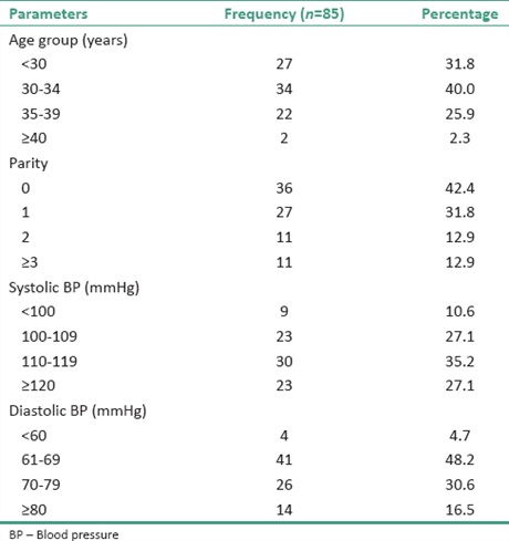
Methodology: This was a prospective longitudinal study in consenting singleton gestation women. The right and the left uterine arteries and the umbilical arteries were interrogated. Doppler parameters; Peak Systolic Velocity (PSV), End Diastolic Velocity (EDV), Resistive Index (RI), Pulsatility Index (PI) and the systolic to diastolic ratio(S/D) were obtained from each healthy pregnant indigenous Nigerian women. Pearson’s correlation analysis of the relationship between these parameters and selected maternal demographic parameters was done. P < 0.05 was considered statistically significant.
Results: The mean of the normal uterine and umbilical arteries values were different from most published reference values
from other parts of the World. No correlation between these indices and maternal parameters were found in this study.
Conclusion: Uterine and umbilical artery Doppler indices among normal indigenous pregnant African women are different from those from the developed World. Using other reference values may be inaccurate for pregnant women in our environment.
Downloads
Article Details
Section

This work is licensed under a Creative Commons Attribution-NonCommercial 4.0 International License.
This is an open access journal, and articles are distributed under the terms of the Creative Commons Attribution-NonCommercial-ShareAlike 4.0 License, which allows others to remix, tweak, and build upon the work non-commercially, as long as appropriate credit is given and the new creations are licensed under the identical terms.
How to Cite
References
1. Betrán AP, Wojdyla D, Posner SF, Gülmezoglu AM. National estimates for maternal mortality: An analysis based on the WHO
systematic review of maternal mortality and morbidity. BMC Public Health 2005;5:131.
2. Olatunji RO, Adekanmi AJ, Obajimi MO, Ojo TO, Roberts OA. Normal ophthalmic artery Doppler velocimetry in healthy pregnant women in Ibadan, South West Nigeria – A preliminary report. West Afr J Ultrasound 2015;11:16‑26.
3. WHO, UNICEF, UNFPA, World Bank Group, and the United Nations Population Division. Trends in Maternal Mortality: 1990 to 2015. Geneva: World Health Organization; 2015. Available from: http://www.who.int/gho/maternal_health/countries/nga.pdf. [Last accessed on 2016 Oct 16].
4. Montague I, Dubbins PA. Clinical applications of Doppler ultrasound in obstetrics. In: Allan P, Dubbins PA, Norman McDicken W, Pozniak MA, editors. Clinical Doppler Ultrasound. 2nd ed. Philadelphia, USA: Churchill Livingstone Elsevier; 2006. p. 315.
5. Lees C, Colin Deane C, Albaiges G. Integrating uterine and fetal Doppler into obstetrics. Making Sense of Obstetrics Doppler
Ultrasound. 1st ed. London: Arnold‑Hodder Headline Group; 2003. p. 53‑64.
6. Aquilina J, Harrington K. Pregnancy hypertension and uterine artery Doppler ultrasound. Curr Opin Obstet Gynecol 1996;8:435‑40.
7. Campbell S, Pearce JM, Hackett G, Cohen‑Overbeek T, Hernandez C. Qualitative assessment of uteroplacental blood flow: Early screening test for high‑risk pregnancies. Obstet Gynecol 1986;68:649‑53.
8. Campbell S, Soothill P. Detection and management of intrauterine growth retardation: A British approach. In: Chervenak FA,
Isaacson GC, Campbell S, editors. Ultrasound in Obstetrics and Gynaecology. Vol. 2. Boston: Little, Brown and Company; 1993.
p. 1431‑5.
9. FitzGerald DE, Drumm JE. Non‑invasive measurement of human fetal circulation using ultrasound: A new method. Br Med J
1977; 2:1450‑1.
10. Campbell S, Diaz‑Recasens J, Griffin DR, Cohen‑Overbeek TE, Pearce JM, Willson K, et al. New Doppler technique for assessing
uteroplacental blood flow. Lancet 1983;1:675‑7.
11. Berkowitz GS, Mehalek KE, Chitkara U, Rosenberg J, Cogswell C, Berkowitz RL. Doppler umbilical velocimetry in the prediction of
adverse outcome in pregnancies at risk for intrauterine growth retardation. Obstet Gynecol 1988;71:742‑6.
12. Bower S, Schuchter K, Campbell S. Doppler ultrasound screening as part of routine antenatal scanning: Prediction of pre‑eclampsia and intrauterine growth retardation. Br J Obstet Gynaecol 1993;100:989‑94.
13. Chanprapaph P, Wanapirak C, Tongsong T. Umbilical artery Doppler waveform indices in normal pregnancies. Thai J Obstet
Gynaecol 2000;12:103‑7.
14. Gómez O, Figueras F, Fernández S, Bennasar M, Martínez JM, Puerto B, et al. Reference ranges for uterine artery mean pulsatility
index at 11‑41 weeks of gestation. Ultrasound Obstet Gynecol 2008;32:128‑32.
15. Lakhkar BN, Ahamed SA. Doppler velocimetry of uterine and umbilical arteries during pregnancy. Indian J Radiol Imaging 1999; 9:119‑25.
16. Kurmanavicius J, Florio I, Wisser J, Hebisch G, Zimmermann R, Müller R, et al. Reference resistance indices of the umbilical, fetal
middle cerebral and uterine arteries at 24‑42 weeks of gestation. Ultrasound Obstet Gynecol 1997;10:112‑20.
17. Acharya G, Wilsgaard T, Berntsen GK, Maltau JM, Kiserud T. Reference ranges for serial measurements of blood velocity and
pulsatility index at the intra‑abdominal portion, and fetal and placental ends of the umbilical artery. Ultrasound Obstet Gynecol
2005;26:162‑9.
18. Arbeille P, Carles G, Bousquet F, Body G, Lansac J. Fetal cerebral and umbilical artery blood flow changes during pregnancy
complicated by malaria. J Ultrasound Med 1998;17:223‑9.
19. Bhide A, Acharya G, Bilardo CM, Brezinka C, Cafici D, Hernandez‑Andrade E, et al. ISUOG practice guidelines: Use of Doppler ultrasonography in obstetrics. Ultrasound Obstet Gynecol 2013;41:233‑39.
20. Griffin JB, Lokomba V, Landis SH, Thorp JM Jr., Herring AH, Tshefu AK, et al. Plasmodium falciparum parasitaemia in the first half of pregnancy, uterine and umbilical artery blood flow, and foetal growth: A longitudinal Doppler ultrasound study. Malar J 2012;
11:319.
21. Hecher K, Campbell S, Doyle P, Harrington K, Nicolaides K. Assessment of fetal compromise by Doppler ultrasound investigation of the fetal circulation. Arterial, intracardiac, and venous blood flow velocity studies. Circulation 1995;91:129‑38.
22. Sutton MS, Theard MA, Bhatia SJ, Plappert T, Saltzman DH, Doubilet P. Changes in placental blood flow in the normal human
fetus with gestational age. Pediatr Res 1990;28:383‑7.
23. Wang Y, Zhao S. Placental blood circulation. Vascular Biology of the Placenta. Ch. 2. San Rafael, CA: Morgan and Claypool Life
Sciences; 2010. Available from: http://www.ncbi.nlm.nih.gov/books/NBK53254/. [Last accessed on 2016 Oct 15].
24. Bahlmann F, Fittschen M, Reinhard I, Wellek S, Steiner E. Reference values for blood flow velocity in the uterine artery in normal
pregnancies from 18 weeks to 42 weeks of gestation calculated by automatic Doppler waveform analysis. Ultraschall Med 2012; 33:258‑64.
25. Peixoto AB, Da Cunha Caldas TM, Tonni G, De Almeida Morelli P, Santos LD, Martins WP, et al. Reference range for uterine artery Doppler pulsatility index using transvaginal ultrasound at 20‑24w6d of gestation in a low‑risk Brazilian population. J Turk Ger Gynecol Assoc 2016;17:16‑20.
26. Oloyede OA, Iketubosin F. Uterine artery Doppler study in second trimester of pregnancy. Pan Afr Med J 2013;15:87.
27. Chanthasenanont A, Nanthakomon T, Pongrojpaw D. Relationship between maternal age and uterine artery Doppler flow during
second trimester. Thai J Obstet Gynaecol 2012;20:173‑8.
28. Pirhonen J, Bergersen TK, Abdlenoor M, Dubiel M, Gudmundsson S. Effect of maternal age on uterine flow impedance. J Clin Ultrasound 2005;33:14‑7.
29. Prefumo F, Bhide A, Sairam S, Penna L, Hollis B, Thilaganathan B. Effect of parity on second‑trimester uterine artery Doppler flow
velocity and waveforms. Ultrasound Obstet Gynecol 2004;23:46‑9.