

Cranial tomographic angiographic evaluation of suspected intracranial vascular abnormalities among a Nigerian cohort
Article Sidebar
Views | PDF/EPUB Downloads:
516
/ 195
/ 35
Main Article Content
Abstract
Background: Lately, there has been an increased utilization of computed tomography angiography (CTA) as the preferred first-line modality for the evaluation and diagnosis of most cerebral vascular lesions.
Objective: The objective of this study was to evaluate suspected intracranial vascular cases, using CTA at a major referral tertiary hospital in South West Nigeria.
Materials and Methods: This was a hospital-based retrospective study of suspected intracranial vascular cases in all ages and both sexes that had CTA from January 2011 to December 2018. Data were analyzed with IBM SPSS version 23.0, and P < 0.005 was considered statistically significant.
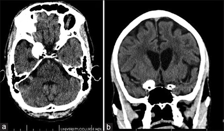
Results: A total of 128 patients were studied, the mean age was 44.1 ± 17.7 years, and male: female ratio was 1:1.06. The leading clinical diagnoses were as follows: intracranial aneurysms (34/128), subarachnoid hemorrhage (27/128), intracranial vascular tumors (26/128), brain hemorrhage from vascular abnormality (19/128), and arteriovenous malformations (AVMs) (10/128). At CTA, 61 patients had vascular abnormalities: intracranial aneurysm was seen in 63.9% with a peak age range of 41–60 years, and the leading location of aneurysms was posterior cerebral artery (18.8%), followed by posterior communicating artery (16.7%) and the cavernous segment of the internal carotid artery (16.7%). AVMs were more common in patients aged 40 years and below (91.7%) in males (66.7%) and in the parietal lobe. Intracranial aneurysms were 3.25 times as common as brain AVMs.
Conclusion: Intracranial aneurysms are the predominant vascular lesions, occurring mostly in the older age group. AVMs occurred mostly in younger people, more in males, and predominantly in the parietal lobes. The hospital incidence of aneurysms to AVMs was 3.25:1.
Downloads
Article Details
Section
This is an open access journal, and articles are distributed under the terms of the Creative Commons Attribution-NonCommercial-ShareAlike 4.0 License, which allows others to remix, tweak, and build upon the work non-commercially, as long as appropriate credit is given and the new creations are licensed under the identical terms.
How to Cite
References
1. Rubin GD, Leipsic J, Schoepf UJ, Fleischmann D, Napel S. CT angiography: A transformation in cardiovascular disease characterization continues to advance. Radiology 2014;271:633‑52.
2. Kalender WA, Seissler W, Klotz E, Vock P. Spiral volumetric CT with single‑breath‑hold technique, continuous transport, and continuous
scanner rotation. Radiology 1990;176:181‑3.
3. Crawford CR, King KF. Computed tomography scanning with simultaneous patient translation. Med Phys 1990;17:967‑82.
4. Alberico RA, Patel M, Casey S, Jacobs B, Maguire W, Decker R. Evaluation of the circle of willis with three‑dimensional CT angiography in patients with suspected intracranial aneurysms. AJNR Am J Neuroradiol 1995;16:1571‑8.
5. Ndubuisi CA, Mezue WC, Ohaegbulam SC, Chikani MC, Ekuma M, Onyia E. Neuroimaging findings in paediatric patients with seizure from an institution in Enugu. Niger J Clin Pract 2016;19:121‑7.
6. Gupta V, Khandelwal N, Prabhakar A, Satish Kumar A, Ahuja CK, Singh P. Prevalence of normal head CT and positive CT findings in a large cohort of patients with chronic headaches. Neuroradiol J 2015;28:421‑5.
7. American Academy of Neurology. Evidence‑Based Guidelines in the Primary Care Setting: Neuroimaging in Patients with Nonacute Headache. Available from: http://tools.aan.com/professionals/practice/pdfs/gl0088.pdf. [Last accessed on 2020 Jul 19].
8. Thomas R, Cook A, Main G, Taylor T, Galizia Caruana E, Swingler R. Primary care access to computed tomography for chronic headache. Br J Gen Pract 2010;60:426‑30.
9. Ezeala‑Adikaibe AB, Ohaegbulam SC, Ndubuisi CA. The Pattern of significant lesions found in computerized tomography scan of recurrent seizure patients at a centre in Enugu, Nigeria. Niger J Clin Pract 2017;20:1289‑93.
10. Feigin VL, Forouzanfar MH, Krishnamurthi R, Mensah GA, Connor M, Bennett DA, et al. Global Burden of Diseases, Injuries, and Risk Factors Study 2010 (GBD 2010) and the GBD Stroke Experts Group. Global and regional burden of stroke during 1990‑2010: findings from the Global Burden of Disease Study 2010. Lancet 2014;383:245‑54. Erratum in: Lancet 2014;383:218.
11. Patel NH, Jain AR, Iyer VK, Shah AG, Jain DA, Shah AA. Clinical – Diagnostic and therapeutic relevance of computed tomography scan of the brain in children with partial seizures. Ann Indian Acad Neurol 2013;16:352‑6.
12. Willems PW, Taeshineetanakul P, Schenk B, Brouwer PA, Terbrugge KG, Krings T. The use of 4D‑CTA in the diagnostic work‑up of brain
arteriovenous malformations. Neuroradiology 2012;54:123‑31.
13. Adeloye AA, Olumide A, Bademosi O, Kolawole TM. Intracranial vascular anomalies in Nigerians. Trop Geogr Med 1981;33:263‑7.
14. Ogbole GI, Ogunseyinde AO, Obajimi MO, Adeyinka OA. Experience with three‑dimensional computed tomographic angiography in Ibadan,
Nigeria. Niger J Clin Pract 2010;13:187‑94.
15. Badr SM, Ahmed Z, Khafaji MA, Alsafi KG, Abbas HY, Jastaniah SD. Computed tomography angiography compared to catheter based angiography in evaluation of cerebral arterial aneurysm and arteriovenous malformation. Open J Med Imaging 2014;4:117‑25.
16. Juvela S, Poussa K, Porras M. Factors affecting formation and growth of intracranial aneurysms: A long‑term follow‑up study. Stroke 2001; 32:485‑91.
17. Backes D, Rinkel GJ, Laban KG, Algra A, Mervyn DI. Vergouwen. patient‑ and aneurysm‑specific risk factors for intracranial aneurysm growth. A systematic review and meta‑analysis. Stroke 2016;47:951‑7.
18. Bokhari MR, Bokhari SR. Brain, Arteriovenous Malformation. In: StatPearls. Treasure Island (FL): StatPearls Publishing; 2018. Available from: https://www.ncbi.nlm.nih.gov/books/NBK430744/. [Last updated on 2017 Apr 26].
19. Singh R, Kumar R, Kumar A. Vascular anomalies of posterior fossa and their implications. J Craniofac Surg 2017;28:2145‑50.
20. Jersey AM, Foster DM. Cerebral aneurysm. In: StatPearls. Treasure Island (FL): StatPearls Publishing; 2020. Available from: https://
www.ncbi.nlm.nih.gov/books/NBK507902/. [Last updated on 2019 Dec 17].
21. Caranci F, Briganti F, Cirillo L, Leonardi M, Muto M. Epidemiology and genetics of intracranial aneurysms. Eur J Radiol 2013;82:1598‑605.
22. The International Study of Unruptured Intracranial Aneurysms Investigators: Unruptured Intracranial Aneurysms‑risk of rupture and risk of surgical intervention. N Engl J Med 1998;339:1725‑33.
23. UCAS Japan Investigators, Morita A, Kirino T, Hashi K, Aoki N, Fukuhara S, et al. The natural course of unruptured cerebral aneurysms in a Japanese cohort. N Engl J Med 2012;366:2474‑82.
24. Jeong YG, Jung YT, Kim MS, Eun CK, Jang SH. Size and location of ruptured intracranial aneurysms. J Korean Neurosurg Soc 2009;45:11‑5.
25. Dehdashti AR, Chiluwal AK, Regli L. The implication of anterior communicating complex rotation and 3‑dimensional computerized tomography angiography findings in surgical approach to anterior communicating artery aneurysms. World Neurosurg 2016;91:34‑42.
26. Badry A, Elshafey R, Khalil M. Detection, characterization, and endovascular therapy planning of intracranial aneurysms with 16‑channel multidetector‑row CT angiography. Egypt J Radiol Nucl Med 2014;45:151‑8.
27. Ergun E, Habera M, Koşar P, Yılmaz A, Koşar U. Diagnostic value of 64‑slice CTA in detection of intracranial aneurysm in patients with SAH and comparison of the CTA results with 2D‑DSA and intraoperative findings. Balkan Med J 2011;28:26‑32.
28. Gamal GH. Diagnostic accuracy of contrast enhancement MRI versus CTA in the diagnosis of intracranial aneurysm in patients with
non‑traumatic subarachnoid hemorrhage. Egypt J Radiol Nucl Med 2015;6:125‑30.
29. Islam M, Chowdhury AH, Msjh C, Islam MN, Nayeem A, Khan SU, et al. A comparative study of computed tomographic angiography and digital subtraction angiography in the evaluation of aneurysmal subarachnoid hemorrhage. J Dhaka med Coll 2013;22:195‑200.
30. Abu Bakar I, Shuaib IL, Mohd Ariff AR, Naing NN, Abdullah JM. Diagnostic cerebral angiography in spontaneous intracranial haemorrhage: A guide for developing countries. Asian J Surg 2005;28:1‑6.
31. Zhu XL, Chan MS, Poon WS. Spontaneous intracranial hemorrhage: Which patients need diagnostic cerebral angiography? A prospective study of 206 cases and review of the literature. Stroke 1997;28:1406‑9.
32. Ferrante L, Acqui M, Trillò G, Lunardi P, Fortuna A. Aneurysms of the posterior cerebral artery: Do they present specific characteristics? Acta Neurochir (Wien) 1996;138:840‑52.
33. Ciceri EF, Klucznik RP, Grossman RG, Rose JE, Mawad ME. Aneurysms of the posterior cerebral artery: Classification and endovascular treatment. AJNR Am J Neuroradiol 2001;22:27‑34.
34. Stapf C, Khaw AV, Sciacca RR, Hofmeister C, Schumacher HC, Pile‑Spellman J, et al. Effect of age on clinical and morphological characteristics in patients with brain arteriovenous malformation. Stroke 2003;34:2664‑9.
35. Milosevic Z. Computed Tomographic Angiography in Intracranial Vascular Diseases; 2000. Available from: https://www.researchgate. net/publication/268347495. [Last accessed on 2020 Jul 19].
36. Bash S, Villablanca JP, Jahan R, Duckwiler G, Tillis M, Kidwell C, et al. Intracranial vascular stenosis and occlusive disease: Evaluation with CT angiography, MR angiography, and digital subtraction angiography. AJNR Am J Neuroradiol 2005;26:1012‑21.
37. Carpenter CR, Hussain AM, Ward MJ, Zipfel GJ, Fowler S, Pines JM, et al. Spontaneous subarachnoid hemorrhage: A systematic review and meta‑analysis describing the diagnostic accuracy of history, physical exam, imaging, and lumbar puncture with an exploration of test thresholds. Acad Emerg Med 2016;23:963‑1003.
38. Westerlaan HE, van Dijk JM, Jansen‑van der Weide MC, de Groot JC, Groen RJ, Mooij JJ, et al. Intracranial aneurysms in patients with subarachnoid hemorrhage: CT angiography as a primary examination tool for diagnosis‑‑systematic review and meta‑analysis. Radiology
2011;258:134‑45. Erratum in: Radiology 2011;260:612.
39. Brouwer PA, Bosman T, van Walderveen MA, Krings T, Leroux AA, Willems PW. Dynamic 320‑section CT angiography in cranial arteriovenous shunting lesions. AJNR Am J Neuroradiol 2010;31:767‑70.
40. Pathan SA, Abosalah S, Nadeem S, Ali A, Hameed AA, Marathe M, et al. Computed tomography abnormalities and epidemiology of adult patients presenting with first seizure to the emergency department in Qatar. Acad Emerg Med 2014;21:1264‑8.
41. Delgado‑Almandoz JE, Schaefer PW, Goldstein JN, Rosand J, Lev MH, González RG, et al. Practical scoring system for the identification of patients with intracerebral haemorrhage at highest risk of harbouring an underlying vascular aetiology: The Secondary Intracerebral Haemorrhage Score. AJNR Am J Neuroradiol 2010;31:1653‑60.
42. Ajiboye N, Chalouhi N, Starke RM, Zanaty M, Bell R. Cerebral arteriovenous malformations: Evaluation and management. Scientific World Journal 2014;2014:649036.
43. Kovač JD, Stanković A, Stanković D, Kovač B, Šaranović D. Intracranial arterial variations: A comprehensive evaluation using CT angiography.
Med Sci Monitor 2014;20:420‑7.
44. Simoni M, Pantoni L, Pracucci G, Palmertz B, Guo X, Gustafson D, et al. Prevalence of CT‑detected cerebral abnormalities in an elderly
Swedish population sample. Acta Neurol Scand 2008;118:260‑7.
45. Schellinger PD. The evolving role of advanced MR imaging as a management tool for adult ischemic stroke: A Western‑European perspective. Neuroimaging Clin N Am 2005;15:245‑58, ix.