

Pattern of Fetal Arterial Blood Flow in selected vessels in Patients With Pregnancy Induced Hypertension in Aminu Kano Teaching Hospital Kano, Nigeria
Article Sidebar
Views | PDF/EPUB Downloads:
60
/ 6
/ 7
Main Article Content
Abstract
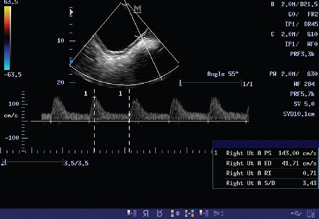
Background: Doppler velocimetry of fetal arterial blood flow in pregnancy induced hypertension (PIH) determines fetal hemodynamic adjustment.
Objective: This study was aimed to determine the pattern of fetal arterial blood flow of selected vessels in patients with PIH.
Materials and Methods: A total of 34 pregnant women with PIH at a gestational age of 24-37 weeks were prospectively examined with Doppler ultrasound of the fetal middle cerebral artery (MCA), umbilical artery and placental blood flow (uterine artery).
Results: The mean peak systolic velocity (PSV) of the fetal MCA was 8.23±3.96, resistance index (RI) was 0.763±0.07 and systolic diastolic (S/D) ratio was 4.558±1.36. The mean PSV of umbilical artery was 72.28±26.585, RI was 0.62±0.19 and S/D was 2.63±0.75. The mean placental blood flow (uterine artery) PSV was 141.34±70.58, RI was 0.59 and S/D was 2.42±1.07. Uterine artery PSV was normal in only six patients. Uterine artery was also not sonographically demonstrated in two patients.
Conclusion: Doppler velocimetry of arterial blood vessels in pregnancy complicated with PIH reveals abnormal pattern; its application in PIH would be useful for further management.
Downloads
Article Details
Section

This work is licensed under a Creative Commons Attribution-NonCommercial 4.0 International License.
This is an open access journal, and articles are distributed under the terms of the Creative Commons Attribution-NonCommercial-ShareAlike 4.0 License, which allows others to remix, tweak, and build upon the work non-commercially, as long as appropriate credit is given and the new creations are licensed under the identical terms.
How to Cite
References
1. Report of the National high blood pressure education program working group on high blood pressure in pregnancy. Am J Obstet
Gynecol 2000;183:S1‑22.
2. Khan KS, Wojdyla D, Say L, Gülmezoglu AM, Van Look PF. WHO analysis of causes of maternal death: A systematic review. Lancet
2006;367:1066‑74.
3. Brosens IA. Morphological changes in the utero‑placental bed in pregnancy hypertension. Clin Obstet Gynaecol 1977;4:573‑93.
4. Dubiel M, Kozber H, Debniak B, Breborowicz GH, Marsal K, Gudmundsson S. Fetal and placental power Doppler imaging in
normal and high‑risk pregnancy. Eur J Ultrasound 1999;9:223‑30.
5. Dubiel M, Gudmundsson S, Gunnarsson G, Marsál K. Middle cerebral artery velocimetry as a predictor of hypoxemia in fetuses with increased resistance to blood flow in the umbilical artery. Early Hum Dev 1997;47:177‑84.
6. Aardema MW, Saro MC, Lander M, De Wolf BT, Oosterhof H, Aarnoudse JG. Second trimester Doppler ultrasound screening of the uterine arteries differentiates between subsequent normal and poor outcomes of hypertensive pregnancy: Two different
pathophysiological entities? Clin Sci (Lond) 2004;106:377‑82.
7. Gupta U, Qureshi A, Samal S. Doppler velocimetry in normal and hypertensive pregnancy. Internet J Gynecol Obstet 2009;11:2.
8. Khalid M, Wahab S, Kumar V, Khalid S, Haroon S, Sabzposh NA. Doppler indices in prediction of fetal outcome in hypertensive
pregnant women. Nepal J Obstet Gynaecol 2011;6:28‑34.
9. Atrash HK, Koonin LM, Lawson HW, Franks AL, Smith JC. Maternal mortality in the United States, 1979‑1986. Obstet Gynecol 1990;76:1055‑60.
10. Miyake H, Nakai A, Koshino T, Araki T. Doppler velocimetry of maternal renal circulation in pregnancy‑induced hypertension. J Clin Ultrasound 2001;29:449‑55.
11. Trudinger BJ, Giles WB, Cook CM. Uteroplacental blood flow velocity‑time waveforms in normal and complicated pregnancy.
Br J Obstet Gynaecol 1985;92:39‑45.
12. Campbell S, Pearce JM, Hackett G, Cohen‑Overbeek T, Hernandez C. Qualitative assessment of uteroplacental blood flow: Early screening test for high‑risk pregnancies. Obstet Gynecol 1986;68:649‑53.
13. Dastur AE, Tank PD. The pharmacology of preventing pre‑eclampsia. J Obstet Gynaecol 2010;60:486‑96.
14. Duckitt K, Harrington D. Risk factors for pre‑eclampsia at antenatal booking: Systematic review of controlled studies. BMJ 2005;330:565.
15. Seely EW, Ecker J. Clinical practice. Chronic hypertension in pregnancy. N Engl J Med 2011;365:439‑46.
16. Jasovic‑Siveska E, Jasovic V, Stoilova S. Previous pregnancy history, parity, maternal age and risk of pregnancy induced hypertension. Bratisl Lek Listy 2011;112:188‑91.
17. Hershkovitz R, Sheiner E, Mazor M. Middle cerebral artery blood flow velocimetry among healthy fetuses with a single umbilical artery. J Ultrasound Med 2006;25:1405‑8.
18. Tarzamni MK, Nezami N, Gatreh‑Samani F, Vahedinia S, Tarzamni M. Doppler waveform indices of fetal middle cerebral artery in normal 20 to 40 weeks pregnancies. Arch Iran Med 2009;12:29‑34.
19. Taslimi MM. Doppler ultrasonography in assessment of fetal well‑being. Neoreviews 2004;5:e247‑51.
20. Lakhkar BN, Ahamed SA. Doppler velocimetry of uterine and umbilical arteries during pregnancy. Genitourin Radiol 1999; 9: 119‑25.