

A comparative control study of ophthalmic artery Doppler velocimetry in patients with primary open angle glaucoma in Kano, Nigeria
Article Sidebar
Views | PDF/EPUB Downloads:
189
/ 32
/ 14
Main Article Content
Abstract
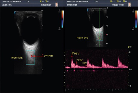
Objective: The objective of the study is to sonographically determine the hemodynamic changes in ophthalmic arteries of patients with primary open angle glaucoma (POAG) at Kano, Nigeria.
Subjects and Methods: We conducted a prospective case–control study at Aminu Kano Teaching Hospital, Nigeria, on 108 newly diagnosed POAG and 108 control subjects. Intraocular pressure (IOP) and Doppler ultrasound velocimetry of ophthalmic arteries were assessed. Peak systolic velocity (PSV), end diastolic velocity (EDV), resistive indices (RIs), pulsatility indices (PIs) and systolic/diastolic (S/D) ratios of the ophthalmic arteries were evaluated and documented.
Results: The mean IOP values of POAG group in the right and left eyes were higher than the values of the right and left eyes of the control group. This was statistically significant (P = 0.000). The mean PSV, EDV, RI, PI, and S/D values in the POAG group of the right and left eyes were lower than values for the right and left eyes of the control group, which was also statistically significant (P = 0.000). The IOP showed positive correlation with PSV and EDV in both eyes of POAG cases but negative correlation with PI and S/D in both eyes in the POAG group. It however correlated positively with RI in the right eye and negatively with RI in the left eye.
Conclusions: The study showed significant differences between ophthalmic artery Doppler indices of patients with POAG and the healthy control subjects.
Downloads
Article Details
Section
This is an open access journal, and articles are distributed under the terms of the Creative Commons Attribution-NonCommercial-ShareAlike 4.0 License, which allows others to remix, tweak, and build upon the work non-commercially, as long as appropriate credit is given and the new creations are licensed under the identical terms.
How to Cite
References
1. Olawoye O, Tarella S. Spectrum of glaucoma presentation in a Nigerian tertiary hospital. Niger J Ophthalmol 2014;22:11‑5.
2. Askira BH, Waziri MA, Musa ZY, Ribadu DY, Kyari FA. Glaucoma awareness among tertiary health care workers in Maiduguri, Nigeria. Bo Med J 2014;2:61‑7.
3. Kyari F, Abdull MM, Wormald R, Evans JR, Nolan W, Murthy GV, et al. Risk factors for open‑angle glaucoma in Nigeria: Results from the Nigeria National Blindness and Visual Impairment Survey. BMC Ophthalmol 2016;16:78.
4. Januleviciene I, Sliesoraityte I, Siesky B, Harris A. Diagnostic compatibility of structural and haemodynamic parameters in open‑angle glaucoma patients. Acta Ophthalmol 2008;86:552‑7.
5. Pinto LA, Vandewalle E, Clerck ED, Marques‑Neves C, Stalmans I. Ophthalmic artery Doppler waveform changes associated with increased damage in glaucoma patients. Invest Ophthalmol Vis Sci 2012;53:2448‑53.
6. Abegão Pinto L, Vandewalle E, Willekens K, Marques‑Neves C, Stalmans I. Ocular pulse amplitude and Doppler waveform analysis in glaucoma patients. Acta Ophthalmol 2014;92:e280‑5.
7. Erickson SJ, Hendrix LE, Massaro BM, Harris GJ, Lewandowski MF, Foley WD et al. Role of colour Doppler imaging in early diagnosis and prediction of progression in glaucoma. Biomed Res Int 2013;2013:871689.
8. Meng N, Zhang P, Huang H, Ma J, Zhang Y, Li H, et al. Color Doppler imaging analysis of retrobulbar blood flow velocities in primary open‑angle glaucomatous eyes: A meta‑analysis. PLoS One 2013;8:e62723.
9. Stalmans I, Vandewalle E, Anderson DR, Costa VP, Frenkel RE, Garhofer G, et al. Use of colour Doppler imaging in ocular blood flow research. Acta Ophthalmol 2011;89:e609‑30.
10. Adeyinka OO, Olugbenga A, Helen OO, Adebayo AV, Rasheed A. Ocular blood flow velocity in primary open angle glaucoma – A tropical African population study. Middle East Afr J Ophthalmol 2013;20:174‑8.
11. Alexandrescu C, Dascalu AM, Mitulescu C, Panca A, Pascu R, Ciuluvica R, et al. Evidence‑based pathophysiology of glaucoma. Maedica (Bucur) 2010;5:207‑13.
12. Srikanth K, Kumar MA, Selvasundari S, Prakash ML. Colour Doppler imaging of ophthalmic artery and central retinal artery in glaucoma patients with and without diabetes mellitus. J Clin Diagn Res 2014;8:C01‑2.
13. Singh MD, Sharma C, Prasad A. A colour Doppler study of retrobulbar blood flow parameters in patients of primary open angle glaucoma. Indian J Clin Exp Ophthalmol 2015;1:84‑90.
14. Budenz DL, Barton K, Whiteside‑de Vos J, Schiffman J, Bandi J, Nolan W, et al. Prevalence of glaucoma in an urban West African population: The Tema Eye Survey. JAMA Ophthalmol 2013;131:651‑8.
15. Olushola O, Oluwatoni O, Omodele J, Anthony B, Gboyega A, Ugochi A, et al. Spectrum of glaucoma presentation in a sub‑urban teaching hospital in South‑Western Nigeria. Health Sci J 2016;10:466.
16. Monsudi KF, Saka ES, Ayodapo AO. Health workers awareness and knowledge of glaucoma in tertiary hospital in Birnin Kebbi, Nigeria. Ophthalmol J 2018;8:1‑8.
17. National Bureau of Statistics. 2015 Statistical Report on Women and Men in Nigeria; 2016. Available from: http://www.national bureau of statistics.gov.ng/downloads. [Last accessed on 2018 Oct 15].
18. Sekeroglu MA, Irkec M, Mocan MC, Ileri E, Dikmenoglu N, Seringec N, et al. The association of ocular blood flow with haemorheological parameters in primary open‑angle and exfoliative glaucoma. Acta Ophthalmol 2011;89:429‑34.
19. Ulickiene R, Paunksnis A. Colour Doppler imaging of the ophthalmic artery in glaucoma patients. Ultragarsas 2004;1:50‑3.
20. Quaranta L, Riva I, Oddone F. 24‑hour IOP fluctuation: Myth or reality. J Mod Ophthalmol 2016;2:103‑9.
21. Kpolovie PJ, Oshodi PO, Iwuchukwu H. Continental inequities in life expectancy. Eur J Biol Med Sci Res 2016;4:30‑47.
22. Kotecha A, Crabb DP, Spratt A, Garway‑Heath DF. The relationship between diurnal variations in intraocular pressure measurements and
central corneal thickness and corneal hysteresis. Invest Ophthalmol Vis Sci 2009;50:4229‑36.
23. Chiou HJ, Chou YH, Liu CJ, Hsu CC, Tiu CM, Teng MM, et al. Evaluation of ocular arterial changes in glaucoma with color Doppler ultrasonography. J Ultrasound Med 1999;18:295‑302.
24. Raut A, Singh M. A comparative study of colour Doppler imaging of ophthalmic artery in primary open angle glaucoma and the age matched healthy volunteers. JMSCR 2017;5:29045‑50.
25. Garhöfer G, Fuchsjäger‑Mayrl G, Vass C, Pemp B, Hommer A, Schmetterer L. Retrobulbar blood flow velocities in open angle glaucoma and their association with mean arterial blood pressure. Invest Ophthalmol Vis Sci 2010;51:6652‑7.
26. Bujang MA, Baharum N. Sample size guideline for correlation analysis. World J Soc Sci Res 2016;3:37‑46.
27. Aggarwal R, Ranganathan P. Common pitfalls in statistical analysis: The use of correlation techniques. Perspect Clin Res 2016;7:187‑90.
28. Sinsomboonthong J. Bias correction in estimation of the population correlation coefficient. Kasetsart J Nat Sci 2013;47:453‑9.
29. Siesky B, Harris A, Racette L, Abassi R, Chandrasekhar K, Tobe LA, et al. Differences in ocular blood flow in glaucoma between patients of African and European descents. J Glaucoma 2015;24:117‑21.
30. Marineta M, Adriana S, Valentin BL, Cristina A. Colour Doppler imaging of the retrobulbar circulation in progressive glaucoma optic neuropathy. Rom J Ophthal 2016;60:237‑48.
31. Eniola MA, Adeyomoye AA, Musa KO, Ishola AA, Olatunji OO. Ophthalmic artery and central retinal artery Doppler patterns in primary open angle glaucoma patients at the Lagos university teaching hospital, Nigeria. J West Afr Coll Surg 2018;8:1‑21.
32. Lekha CS, Mini PA, Josey VT. Doppler evaluation of ocular vessels in primary open angle glaucoma patients. JMSCR 2017;5:27907‑16.
33. Asejczyk‑Widlicka M, Krzyzanowska‑Berkowska P, Sander BP, Iskander DR. Age‑related changes in ocular blood velocity in suspects
with glaucomatous optic disc appearance. Comparison with healthy subjects and glaucoma patients. PLoS One 2015;10:e0134357.
34. Abegão Pinto L, Vandewalle E, Stalmans I. Disturbed correlation between arterial resistance and pulsatility in glaucoma patients. Acta Ophthalmol 2012;90:e214‑20.
35. Calvo P, Ferreras A, Polo V, Güerri N, Seral P, Fuertes‑Lazaro I, et al. Predictive value of retrobulbar blood flow velocities in glaucoma
suspects. Invest Ophthalmol Vis Sci 2012;53:3875‑84.