

Sonographic Triad In Abdominal Pregnancy: Illustration With Four Cases
Article Sidebar
Views | PDF/EPUB Downloads:
227
/ 37
Main Article Content
Abstract
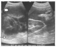
Background: Abdominal pregnancy (AP) is a rare form of ectopic gestation with a high morbidity and mortality. The diagnosis is difficult because of its relatively non-specific asymptomatic nature and non-specificity of symptoms. Ultrasonography may be the first pointer to the diagnosis.
METHODS: Four cases of AP diagnosed between 16-38weeks gestation and had laparotomy confirmations are presented. Focused obstetrics ultrasound scans, which sought to identify the upper vagina, cervix and uterus assisted our diagnosis.
RESULTS: Our first case of AP was missed at ultrasonography. Thereafter, three consecutive cases were correctly diagnosed. Only one out of our four patients had the pregnancy delivered of live baby. The unique sonographic features common to all are triad of masses: the empty uterus, the placenta and the closely packed bowel loops with omentum.
CONCLUSIONS: In obstetrics scan, the finding of unusual lower intra-abdominal masses during panoramic scans should arouse Sonologists or Sonographers to suspect AP until rigorous and lucid pelvic ultrasound scans that must have identified the upper vagina, cervix and the uterus prove otherwise.
Downloads
Article Details
Section

This work is licensed under a Creative Commons Attribution-NonCommercial 4.0 International License.
This is an open access journal, and articles are distributed under the terms of the Creative Commons Attribution-NonCommercial-ShareAlike 4.0 License, which allows others to remix, tweak, and build upon the work non-commercially, as long as appropriate credit is given and the new creations are licensed under the identical terms.
How to Cite
References
1. Stanley JH, Horger EO 3rd, Fagan CJ, Andriole JG, Fleischer AC. Sonographic findings in abdominal pregnancy. AJR Am J Roentgenol. 1986;147:1043-6.
2. Abdulkadir AY, Aboyeji PA, Adesiyun OAM, Fawole AA. Abdominal pregnancy presenting with recurrent severe anaemia and massive retroplacental haemorrhage. Sexual Health Matters. 2008;9:14-17.
3. Ayinde OA, Aimakhu CO, Adeyanju OA, Omigbodun AO. Abdominal pregnancy at the University College Hospital, Ibadan: A ten-year review. Afr J Reprod Health. 2005;9:123-7.
4. Pricop M, Tomosoiu C, Feurdean M, Gafencu C, Musca S, Slatineanu S, Daniil C. Abdominal pregnancy. Case report, review of the literature. Rev Med Chir Soc Med Nat Iasi. 2000;104:139-42.
5. Angtuaco TL, Shah HR, Neal MR, Quirk JG. Ultrasound evaluation of abdominal pregnancy. Crit Rev Diagn Imaging. 1994;35(1):1-59.
6. Allibone GW, Fagan CJ, Porter SC. The sonographic features of intra-abdominal pregnancy. J Clin Ultrasound. 1981; 9:383-7.
7. Graham D, Johnson TR Jr, Sanders RC. Sonographic findings in abdominal pregnancy. JUltrasound Med. 1982; 1:71-4.
8. Ali V, Saldana LR, Balat IY, Katragadda R. Pitfalls in sonographic diagnosis of abdominal pregnancy. South Med J. 1981;74:477-9.
9. Spanta R, Roffman LE, Grissom TJ, Newland JR, McManus BM. Abdominal pregnancy: magnetic resonance identification with ultrasonographic follow-up of placental involution. Am J Obstet Gynecol. 1987; 15:887-9.
10. Lockhat F, Corr P, Ramphal S, Moodley J. The value of magnetic resonance imaging in the diagnosis and management of extra-uterine abdominal pregnancy. Clin Radiol. 2006;61:264-9.
11. Gerli S, Rossetti D, Baiocchi G et al. Early ultrasonographic diagnosis and laparoscopic treatment of abdominal pregnancy. Eur J Obstet Gynecol Reprod Biol. 2004; 15:103-5.
12. Sepúlveda WH. Sonographic diagnosis of combined intrauterine and extrauterine pregnancy. Int J Gynaecol Obstet. 1990;31: 361-4.