

Doppler ultrasound evaluation of blood flow patterns of the uterine arteries in pre‑ and postmenopausal women with cervical cancer and controls in Zaria
Article Sidebar
Views | PDF/EPUB Downloads:
367
/ 65
/ 23
Main Article Content
Abstract
Background: Cervical cancer remains an important health issue, especially in the developing countries that account for about 85% of the world burden of cervical cancer. Finding a role for Doppler ultrasound in the evaluation of these patients may reduce the cost and improve access to management.
Aim: The study aimed at evaluating the Doppler flow parameters in pre- and postmenopausal patients with cervical cancer when compared to normal controls in a teaching hospital in Nigeria.
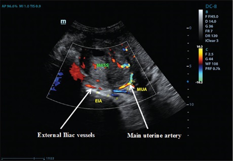
Methodology: This is a prospective case–control observational study conducted over a period of 7 months (August 2016–February 2017) in Ahmadu Bello University Teaching Hospital, Zaria, Nigeria. Eighty-one patients with cervical cancer and 81 age-matched controls had a transabdominal Doppler ultrasound examination of the main uterine arteries. The data were analyzed using SPSS version 20.0, Chicago, Illinois, USA. Statistical differences in the uterine artery indices between two groups were tested, and P < 0.05 was considered as statistically significant.
Results: The mean resistive index (RI) and pulsatility index (PI) of the patients were 0.64 ± 0.12 and 1.26 ± 0.31, respectively, and of controls were 0.88 ± 0.08 and 2.60 ± 0.56, respectively. These showed significantly lower values in patients than the controls (P < 0.0001). The mean end-diastolic velocity (EDV) was significantly higher in patients than the controls (P < 0.0001). There was, however,
no significant difference in the mean peak systolic velocity (PSV) in patients and controls (P = 0.97). Both premenopausal patients and controls had significantly lower RI and PI and significantly higher PSV and EDV compared to their postmenopausal counterpart.
Conclusion: The findings showed that significant differences exist in the uterine artery Doppler flow parameters in patients with cervical cancer compared to the healthy controls and that these parameters are influenced by menopausal status of the women and the size of the cervical mass. Hence, Doppler helps in staging, prognosticating, and posttreatment evaluation of patients with cervical cancer.
Downloads
Article Details
Section
This is an open access journal, and articles are distributed under the terms of the Creative Commons Attribution-NonCommercial-ShareAlike 4.0 License, which allows others to remix, tweak, and build upon the work non-commercially, as long as appropriate credit is given and the new creations are licensed under the identical terms.
How to Cite
References
1. World Health Organization. Comprehensive Cervical Cancer Control. Available from: http://www.who.int/reproductivehealth/
publications/cancers/cervicalcancer‑guide/en/. [Last assessed on 2015 Oct 25].
2. Ferlay J, Shin HR, Bray F, Forman D, Mathers C, Parkin DM. Estimates of worldwide burden of cancer in 2008: GLOBOCAN 2008. Int J
Cancer 2010;127:2893‑917.
3. Walboomers JM, Jacobs MV, Manos MM, Bosch FX, Kummer JA, Shah KV, et al. Human papillomavirus is a necessary cause of invasive
cervical cancer worldwide. J Pathol 1999;189:12‑9.
4. World Health Organization ICO. Information Centre of Human Papilloma Virus and Cervical Cancer: Human papilloma virus and related cancers in the world. Summary Report. World Health Organization ICO; 2010. Available from: http://www.who.int/hpvcentre/en. [Last accessed on 2015 Jun 20].
5. Willoughby BJ, Faulkner K, Stamp EC, Whitaker CJ. A descriptive study of the decline in cervical screening coverage rates in the North East and Yorkshire and the Humber regions of the UK from 1995 to 2005. J Public Health (Oxf) 2006;28:355‑60.
6. KyariO, NggadaH, MairigaA. Malignant tumours of female genital tract in North Eastern Nigeria. East Afr Med J 2004;81:142‑5.
7. Uzoigwe SA, Seleye‑Fubara D. Cancer of the uterine cervix in Port Harcourt, rivers state: A clinic‑pathological review. Niger J Med 2004;13:110‑3.
8. Yakasai IA, Ugwa EA, Otubu J. Gynaecological malignancies in Aminu Kano teaching hospital Kano: A 3 year review. Niger J Clin Pract
2013;16:63‑6.
9. Adewole IF, Edozien LC, Babarinsa IA, Akang EE. Invasive and in situ carcinoma of the cervix in young Nigerians: A clinico‑pathologic study
of 27 cases. Afr J Med Med Sci 1997;26:191‑3.
10. Folkman J, Watson K, Ingler D, Hanahan D. Introduction of angiogenesis during the transition from hyperplasia to neoplasia. Nature 1989;339:58‑61.
11. Kidron D, Bernheim J, Aviram R, Cohen I, Fishman A, Beyth Y, et al. Resistance to blood flow in ovarian tumors: Correlation between resistance index and histological pattern of vascularization. Ultrasound Obstet Gynecol 1999;13:425‑30.
12. Kaku T, Hirakawa T, Kamura T, Amada S, Kinukawa N, Kobayashi H, et al. Angiogenesis in adenocarcinoma of the uterine cervix. Cancer 1998;83:1384‑90.
13. Abulafia O, Sherer DM. Angiogenesis in the uterine cervix. Int J Gynecol Cancer 2000;10:349‑57.
14. Obermair A, Wanner C, Bilgi S, Speiser P, Kaider A, Reinthaller A. Tumour angiogenesis in stage 1B cervical cancer: Correllation of microvessel density with survival. AM J Obstet Gynaecol 1998;178:314‑9.
15. Dinh TV, Hannigan EV, Smith ER, Hove MJ, Chopra V, To T. Tumour angiogenesis as predictor of recurrence in stage 1b squamous cell carcinoma of the cervix. Gynecol Oncol 1996;87:751‑4.
16. Taylor KJ, Ramos I, Carter D, Morse SS, Snower D, Fortune K. Correlation of Doppler US tumor signals with neovascular morphologic features. Radiology 1988;166:57‑62.
17. Maulik D, Ivica Z. Doppler in Obstetrics and Gynaecology. 2nd ed. New York‑USA: Springer‑Verlage; 2005. p. 227‑42.
18. Follen M, Levenback CF, Lyer RB, Grisgsby PW, Boss EA, DelpassandES, et al. Imaging in cervical cancer. Cancer 2003;98:2028‑38.
19. Choi HJ, Ju W, Myung SK, Kim Y. Diagnostic performance of computer tomography, magnetic resonance imaging, and positron
emission tomography or positron emission tomography/computer tomography for detection of metastatic lymph nodes in patients with
cervical cancer: Meta‑analysis. Cancer Sci 2010;101:1471‑9.
20. Liyanage SH, Roberts CA, Rockall AG. MRI and PET scans for primary staging and detection of cervical cancer recurrence. Womens Health (Lond) 2010;6:251‑67.
21. McDicken WN, Hoskins PR. Physics: Principle, practice and artifacts. In: Pozniak AM, Allan PL, editors. Clinical Doppler Ultrasound. 3rd ed.
London, UK: Churchill Livinstone; 2014. p. 1‑25.
22. Ochi H, Suginami H, Matsubara K, Taniguchi H, Yano J, Matsuura S. Micro‑bead embolization of uterine spiral arteries and changes in uterine arterial flow velocity waveforms in the pregnant ewe. Ultrasound Obstet Gynecol 1995;6:272‑6.
23. Maulik D, Mundy D, Heitmann E, Maulik D. Evidence‑based approach to umbilical artery Doppler fetal surveillance in high‑risk pregnancies: An update. Clin Obstet Gynecol 2010;53:869‑78.
24. National Population Commission. National Demographic Survey. National Population Commission; 2006. Available from: http://www.
population.gov.ng. [Last assessed on 2015 Jun 04].
25. Araoye MO. Research Methodology with Statistics for Health and Social Scieces. Ilorin‑Nigeria: Nathadex Publishers; 2003. p. 115‑22.
26. Durowade KA, Osagbemi GK, Salaudeen AG, Musa OI, Akande TM, Babatunde OA, et al. Prevalence and risk factors of cervical cancer
among women in an urban community of Kwara State, North central Nigeria. J Prev Med Hyg 2012;53:213‑9.
27. Bhide A, Acharya G, Bilardo CM, Brezinka C, Cafici D, Hernandez‑Andrade E, et al. ISUOG practice guidelines: Use of Doppler ultrasonography in obstetrics. Ultrasound Obstet Gynecol 2013;41:233‑39.
28. Sule ST, Shehu MS. Cervical cancer management in Zaria, Nigeria. Afr J Health Sci 2007;14:149‑53.
29. Ijaiya MA, Aboyeji PA, Buhari MO. Cancer of the cervix in Ilorin, Nigeria. West Afr J Med 2004;23:319‑22.
30. Anorlu RI, Orakwue CO, Oyeneyin L, Abudu OO. Late presentation of patients with cervical cancer to a tertiary hospital in Lagos: What is responsible? Eur J Gynaecol Oncol 2004;25:729‑32.
31. Abdul MA, Shittu SO, Mohammed A, Mayun A. Non‑squamous cell carcinoma of the cervix in Zaria, Northern Nigeria: A clinicopathological analysis. Ann Afr Med 2006;5:118‑21.
32. Egbang NJ, Tchente NC, Owona ML, Simo G, Essam SJ, Elono FA, et al. Epidemiological and histological profile of cervical cancer in
Cameroon: About 2078 cases. Open J Obstet Gynecol 2016;6:232‑9.
33. Pérez‑Gómez B, Martínez C, Navarro C, Franch P, Galceran J, Marcos‑Gragera R, et al. The moderate decrease in invasive cervical cancer incidence rates in Spain (1980‑2004): Limited success of opportunistic screening? Ann Oncol 2010;21 Suppl 3:iii61‑8.
34. Blood CH, Zetter BR. Tumor interactions with the vasculature: Angiogenesis and tumor metastasis. Biochim Biophys Acta 1990; 1032: 89‑118.
35. McDonald DM, Choyke PL. Imaging of angiogenesis: From microscope to clinic. Nat Med 2003;9:713‑25.
36. Di Vagno G, Cormio G, Vimercati A, Nacci G, Greco P, Lepera A, et al. Transvaginal color‑Doppler sonography for monitoring the response
to neoadjuvant chemotherapy in patients with locally advanced cervical cancer. Minerva Ginecol 1996;48:463‑7.
37. Greco P, Cormio G, Vimercati A, Loverro G, Selvaggi L. Transvaginal colour Doppler sonography In predicting the response to chemotherapy in advanced cervical carcinoma. Ultrasound Obstet Gynecol 1997;9:49‑52.
38. Belitsos P, Papoutsis D, Rodolakis A, Mesogitis S, Antsaklis A. Three‑dimensional power Doppler ultrasound for the study of cervical cancer and precancerous lesions. Ultrasound Obstet Gynecol 2012;40:576‑81.
39. Testa AC, Ferrandina G, Distefano M, Fruscella E, Mansueto D, Basso D, et al. Color Doppler velocimetry and three‑dimensional color power angiography of cervical carcinoma. Ultrasound Obstet Gynecol 2004;24:445‑52.
40. Bolla D, In‑Albon S, Papadia A, Di Naro E, Gasparri ML, Mueller MM, et al. Doppler ultrasound flow evaluation of the uterine arteries significantly correlates with tumor size in cervical cancer patients. Ann Surg Oncol 2015;22 Suppl 3:S959‑63.
41. LuziG, CoataG, CucchiaGC, CosmiEV, Di RenzoGC. Doppler studies of uterine arteries in spontaneous and artificially induced menopausal women. Ultrasound Obstet Gynecol 1993;3:354‑6.
42. Cheng WF, Lee CN, Chu JS, Chen CA, Chen TM, Shau WY, et al. Vascularity index as a novel parameter for the in vivo assessment of angiogenesis