

Magnetic Resonance Imaging of Lumbosacral Intervertebral Discs in Nigerians with Low Back Pain
Article Sidebar
Views | PDF/EPUB Downloads:
78
/ 12
/ 7
Main Article Content
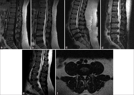
Abstract
Background: Magnetic resonance imaging (MRI) is the modality of choice in diagnostic imaging of the neurological structures related to low back pain (LBP). Particularly, in evaluation of the vertebral discs, its relationship to the nerve roots and related structures with precise diagnosis that guides the patient’s management. This study evaluated the spectrum of intervertebral disc findings in LBP patients at two imaging centers in the South-West and North-Central Nigeria. Association between the clinical diagnosis and MRI disc findings was tested.
Materials and Methods: This is a retrospective descriptive study. The request cards, reports, and available
recorded images of patients referred for MRI for LBP from 2013 till 2015 were retrieved. The extracted patients’ information, radiological findings were documented in a data form, with due compliance to confidentiality of the cohort and analyzed by Statistical Package for Social Sciences (SPSS) software (Version 20.0., IBM Corp. Released 2011, IBM SPSS Statistics for Windows, IBM Corp. Armonk, NY, USA) Results were presented as tables and test of association between variables carried out using Pearson t-test.
Results: A total number of 205 patients were enrolled. Age range of the subjects was 10–83 years and mean age = 52.5 ± 15.4 years. There were more females with a male to female ratio of 1:1.04. LBP was more common in the fifth decade and least in ≤20 years (3%). MRI disc abnormalities increased with advancing age with statistically significant association between disc abnormalities and the patient’s age, lumbar spondylosis, disc prolapse, and spinal canal stenosis.
Conclusion: Disc abnormalities increased significantly with advancing age. Lumbar spondylosis, disc prolapse, and spinal canal stenosis are most commonly associated with florid disc abnormalities on MRI.
Downloads
Article Details
Section

This work is licensed under a Creative Commons Attribution-NonCommercial 4.0 International License.
This is an open access journal, and articles are distributed under the terms of the Creative Commons Attribution-NonCommercial-ShareAlike 4.0 License, which allows others to remix, tweak, and build upon the work non-commercially, as long as appropriate credit is given and the new creations are licensed under the identical terms.
How to Cite
References
1. Andersson GB. Epidemiological features of chronic low‑back pain. Lancet 1999;354:581‑5.
2. Hart LG, Deyo RA, Cherkin DC. Physician office visits for low back pain. Frequency, clinical evaluation, and treatment patterns from a U.S. national survey. Spine (Phila Pa 1976) 1995;20:11‑9.
3. Deyo RA, Weinstein J. Primary health – Low back pain. N Engl J Med 2001;344:363‑70.
4. Ross JS, Modic MT, current assessment of spinal degenerative disease with magnetic resonance imaging. Cli ortho Rel Res 1992; (279):68‑81.
5. Ebbehøj NE, Hansen FR, Harreby MS, Lassen CF. Low back pain in children and adolescents. Prevalence, risk factors and prevention. Ugeskr Laeger 2002;164:755‑8.
6. Harreby M, Nygaard B, Jessen T, Larsen E, Storr‑Paulsen A, Lindahl A, et al. Risk factors for low back pain in a cohort of 1389
Danish school children: An epidemiologic study. Eur Spine J 1999;8:444‑50.
7. Watson KD, Papageorgiou AC, Jones GT, Taylor S, Symmons DP, Silman AJ, et al. Low back pain in schoolchildren: Occurrence and
characteristics. Pain 2002;97:87‑92.
8. Balagué F, Skovron ML, Nordin M, Dutoit G, Pol LR, Waldburger M. Low back pain in schoolchildren. A study of familial and
psychological factors. Spine (Phila Pa 1976) 1995;20:1265‑70.
9. Taimela S, Kujala UM, Salminen JJ, Viljanen T. The prevalence of low back pain among children and adolescents. A nationwide,
cohort‑based questionnaire survey in Finland. Spine (Phila Pa 1976) 1997;22:1132‑6.
10. Leboeuf‑Yde C, Kyvik KO. At what age does low back pain become a common problem? A study of 29,424 individuals aged 12‑41 years. Spine (Phila Pa 1976) 1998;23:228‑34.
11. Merskey H, Bogduk N, editors. Classification of Chronic Pain. 2nd ed. Seattle, USA: IASP Press; 1994.
12. Brown MD. The source of low back pain and sciatica. Seminars in Arthritis and Rheumatism 1989;18:4(Suppl 2):67‑72. Available from: http://www.sciencedirect.com/science/journal/00490172/18/4/supp/S2.
13. Cassidy JD, Carroll LJ, Côté P. The Saskatchewan health and back pain survey. The prevalence of low back pain and related disability in Saskatchewan adults. Spine (Phila Pa 1976) 1998;23:1860‑6.
14. Bigos SJ, Battié MC, Spengler DM, Fisher LD, Fordyce WE, Hansson TH, et al. A prospective study of work perceptions and
psychosocial factors affecting the report of back injury. Spine (Phila Pa 1976) 1991;16:1‑6. [Erratum in: Spine 1991;16:688].
15. Papageorgiou AC, Rigby AS. Review of UK data on the rheumatic diseases – 7. Low back pain. Br J Rheumatol 1991;30:208‑10.
16. Young PY, Alies NA, Shuaib IL. Correlation of clinical presentation, radiography and magnetic resonance imaging for low back pain in a preliminary survey. J HK Coll Radiol 2003;6:144‑57.
17. Humphreys SC, Eck JC, Hodges SD. Neuroimaging in low back pain. Am Fam Physician 2002;65:2299‑306.
18. Ito M, Incorvaia KM, Yu SF, Fredrickson BE, Yuan HA, Rosenbaum AE. Predictive signs of discogenic lumbar pain on magnetic resonance imaging with discography correlation. Spine (Phila Pa 1976) 1998;23:1252‑8.
19. Kjaer P, Leboeuf‑Yde C, Sorensen JS, Bendix T. An epidemiologic study of MRI and low back pain in 13‑year‑old children. Spine (Phila Pa 1976) 2005;30:798‑806.
20. Kjaer P. Low back pain in relation to lumbar spine abnormalities
as identified by magnetic resonance imaging. Dan Med Bull
2005;52:30.
21. Irurhe NK, Adekola OO, Quadri AR, Menkiti ID, Udenze IC, Awolola NA. The magnetic resonance scan findings in adult Nigerian with low back pain. World J Med Sci 2012;7:204‑9.
22. Adeyinka AO, Omidiji OA. Magnetic resonance imaging diagnoses in the lumbar spine of adults with low back pain in South West
Nigeria. West Afr J Radiol 2011;18:1‑9.
23. Mustapha Z, Ahmadu MS, Ali AA, Ibrahim K, Okedayo M. Patterns of requests and findings in magnetic resonance imaging (MRI) of the lumbosacral spine at University of Maiduguri Teaching Hospital, Northeastern Nigeria. IOSR J Dent Med Sci 2013;11:18‑24.
24. Omoke NI, Amaraegbulam PI. Low back pain as seen in orthopedic clinics of a Nigerian Teaching Hospital. Niger J Clin Pract
2016;19:212‑7.
25. Galukande M, Muwazi S, Mugisa DB. Aetiology of low back pain in Mulago Hospital, Uganda. Afr Health Sci 2005;5:164‑7.
26. Uduma FU, Ongolo P, Assam G, Fokam P, Motah M. Evaluation of pattern of magnetic resonance images of lumbosacral spine in
Cameroon. A Pioneer study. Glob J Med Res 2011;11:30‑41.
27. Orege JA, Abuya JM, Elias GD. Common magnetic resonance imaging (MRI) patterns in patients with low back pain in Eldoret,
Kenya. J Sci Innov Res 2013;2:260‑79.
28. Aprill C, Bogduk N. High‑intensity zone: A diagnostic sign of painful lumbar disc on magnetic resonance imaging. Br J Radiol
1992;65:361‑9.
29. Greenspan A. Orthopaedic imaging: A practical approach. 4th ed. Philadelphia, PA: Lippincott Williams & Wilkins; 2004. p. 459.
30. Cassar‑Pullicino VN. MRI of the ageing and herniating intervertebral disc. Eur J Radiol 1998;27:214‑28.
31. Rehman L, Khaleeq S, Hussain A, Mushtaq GE, Zaman K. Correlation between clinical features and magnetic resonance imaging findings in patients with lumbar disc herniation. J Postgrad Med Inst 2007;21:65‑70.
32. Jensen MC, Brant‑Zawadzki MN, Obuchowski N, Modic MT, Malkasian D, Ross JS. Magnetic resonance imaging of the lumbar
spine in people without back pain. N Engl J Med 1994;331:69‑73.
33. DeCandido P, Reinig JW, Dwyer AJ, Thompson KJ, Ducker TB. Magnetic resonance assessment of the distribution of lumbar spine
disc degenerative changes. J Spinal Disord 1988;1:9‑15.