

Renal Complications of sickle Cell Anemia in Zaria, Nigeria: An ultrasonographic Assessment
Article Sidebar
Views | PDF/EPUB Downloads:
141
/ 11
/ 12
Main Article Content
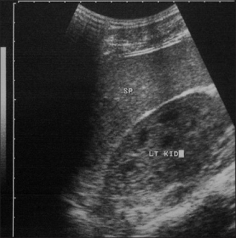
Abstract
Background: Vaso-occlusion in the kidney is a capillary phenomenon. Renal medullary hyperosmolalrity and low oxygen tension
encourage the maximum formation of sickled red cells. In addition, the glomerular loops constitute an aggregating point for the sickled red cells with subsequent obstruction to the blood flow. These factors, together with the large volume of blood flowing through the kidneys make renal complications inevitable in sickle cell anemia (SCA).
Aim: The purpose of this prospective study was to report the renal sonographic findings of a sample of patients with hemoglobin electrophoretic pattern consistent with sickle cell anemia
Materials and Methods: A cross-sectional prospective study of 74 patients with the diagnosis of SCA, as documented by electrophoresis and who attended the adult sickle cell clinic, Hematology Department of the Ahmadu Bello University Teaching Hospital, Zaria, Nigeria and 20 age-matched controls with a normal hemoglobin (HbAA) phenotype and with no history of renal disease, was carried out between April and December, 2010. None of the patients had any clinical evidence of acute sickle episode (crisis) at the time of ultrasonographic examination. B-mode ultrasonography with Aloka SSD-3500 was used to assess the kidneys. The hematological parameters were determined by multiparameter analyzer Sysmex XT 2000i, whereas creatinine and urea of the patients were also analyzed using the Selectra XL chemistry autoanalyzer. results: Renal size of the study group was compared with that of the control group and it showed a significant increase in the adult patients with SCA (P<0.05). The mean right renal length in the study group and control group was 10.65±0.97 cm and 9.95±0.80 cm (P<0.001), respectively, whereas the mean left renal length in the study group and control group was 10.70±1.02 cm and 10.00±0.66 cm (P<0.001), respectively. Statistical relationships between renal length and some hematological indices (packed cell volume (PCV), red blood cell count, and reticulocyte count) showed no correlation but renal length was positively correlated with reticulocyte count, especially high reticulocyte count. Other findings documented and discussed include echogenicity of renal parenchyma, hydronephrosis, and papillary necrosis.
Conclusion: Renal ultrasound imaging of patients with SCA showed a high incidence of renal abnormalities.
Downloads
Article Details
Section

This work is licensed under a Creative Commons Attribution-NonCommercial 4.0 International License.
This is an open access journal, and articles are distributed under the terms of the Creative Commons Attribution-NonCommercial-ShareAlike 4.0 License, which allows others to remix, tweak, and build upon the work non-commercially, as long as appropriate credit is given and the new creations are licensed under the identical terms.
How to Cite
References
1. Herrick JB. Peculiar elongated and sickle‑shape red blood corpuscles in a case of severe anemia. Arch Int Med 1910;6:517.
2. Sydenstricker VP, Mulherin WA, Houseal RW. Sickle cell anemia, report of 2 cases in children with necropsy in one case. Am J Dis Child 1923; 26:132.
3. Konotey‑Ahula FI. The sickle cell diseases. Arch Intern Med 1974;133:611.
4. Lonergan GJ, Cline DB, Abbondanzo SI. From the Archives of the AFIP: Sickle cell anemia. Radiographics 2001;21:971‑4.
5. Fixer J, Styles L. Sickle cell disease. Pediatr Clin North Am 2002;49:1193‑210.
6. Papadaki MG, Kattamis AC, Paapadaki IG. Abdominal ultrasonographic findings in patients with sickle cell anemia and thalassemia
intermedia. Pediatr Radiol 2003;33:515‑21.
7. Van‑Eps LW, de Jong. Sickle cell disease. In: Diseases of the kidney, Schrier RW, Gottschalk CW, editors. Boston: Little, Brown and Co.; 1997. p. 561‑90.
8. Karen L, Gardiner RT. The Renal Sonographic Appearance of Sickle Cell Disease. Med J of Ultra. 1987;3:14‑9.
9. Walker TM, Beardsall K, Thomas PW, Serjeant GR. Renal length in sickle cell disease: Observations from a cohort study. Clin Nephrol
1996;46:384‑8.
10. Balci A, Karazincir S, Sangün O, Gali E, Daplan T, Cingiz C, et al. Prevalence of abdominal ultrasonographic abnormalities in patients with sickle cell disease. Diagn Interv Radiol 2008;14:133‑7.
11. Mapp E, Karasick S, Pollack H, Wechsler RJ, Karasick D. Uroradiological manifestations of S‑hemoglobinopathy. Semin Roentgenol 1987; 22:186‑94.
12. Goodman MS, Jacobs JA. Sickle cell Hematuria controlled by intrarenal oxychlorosene irrigation. J Urol 1983;130:326‑7.
13. Ataga KI, Orringer EP. Renal abnormalities in sickle cell disease. Am J Hematol 2000;63:205‑11.
14. Zinn D, Haller JO, Cohen HL. Focal and diffuse increased echogenicity in the renal parenchyma in patient with sickle hemoglobinopathies: An observation. J Ultra Med 1993;12:211‑4.
15. Namjoshi SP. Punctate echogenic foci in spleen and increased echogenicity in renal cortex in sickle cell anemia. J Clin Ultrasound 1999; 27:52.
16. Shultz PK, Strife JL, Strife CF, McDaniel JD. Hyperechoic renal medullary pyramids in infants and children. Radiology 1991; 181:163‑7.
17. Hoffman JC, Schnur MJ, Koenigsberg M. Demonstration of renal papillary necrosis by sonography. Radiology 1982;145:785‑7.