

Delayed Superficial Femoral Artery Pseudoaneurysm Following Penetrating Trauma in a Young Patient with Sonographic Findings
Article Sidebar
Views | PDF/EPUB Downloads:
171
/ 17
/ 11
Main Article Content
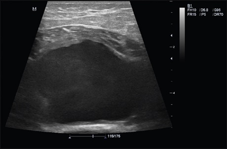
Abstract
The most common causes of penetrating arterial injuries are stab and gunshot-related injuries. Any penetrating trauma to the
vessel wall that causes damage to the arterial wall will result in a pseudoaneurysm. The time from initial injury to detection of the
pseudoaneurysm has been reported to vary from hours to years, depending on the site of formation and clinical symptoms. Enlarging
swelling, the presence of pulsatile mass, palpable thrill, edema, and paresthesia of the involved area can be present based on location.
Ultrasonography (US) and color Doppler US have been the preferred initial imaging technique to evaluate the vascular structures,
especially under emergency conditions. The detection of a turbulent flow that appears as a classic “yin‑yang” sign is a characteristic
feature of pseudoaneurysms on the color Doppler US. In addition, the identification of a “to and fro” spectral waveform in the neck is
considered pathognomonic for a pseudoaneurysm. As per the literature, the color Doppler US demonstrated high sensitivity (94%)
and specificity (97%) for the diagnosis of a pseudoaneurysm. Therefore, it is a noninvasive, inexpensive, easy, and very tolerable first
choice method for the diagnosis of a pseudoaneurysm. Here, we report on a delayed posttraumatic distal superficial femoral artery
pseudoaneurysm in a young patient with color Doppler US findings.
Downloads
Article Details
Section

This work is licensed under a Creative Commons Attribution-NonCommercial 4.0 International License.
This is an open access journal, and articles are distributed under the terms of the Creative Commons Attribution-NonCommercial-ShareAlike 4.0 License, which allows others to remix, tweak, and build upon the work non-commercially, as long as appropriate credit is given and the new creations are licensed under the identical terms.
How to Cite
References
1. Sharma S, Bhargava B, Mahapatra M, Malhotra R. Pseudoaneurysm of the superficial femoral artery following accidental trauma: Result of treatment by percutaneous stent‑graft placement. Eur Radiol 1999;9:422‑4.
2. Kouvelos GN, Papa N, Matsagkas MI. Spontaneous superficial femoral artery giant false aneurysm. ANZ J Surg 2011;81:655‑6.
3. Alsmady MM, Abdallah FF, Shanti HA, Samara OM. Spontaneous femoral artery pseudoaneurysm in a young patient. Ann Vasc Surg 2013; 27:972.e7‑9.
4. Schena S, Owens CA, Hassoun HT, Kibbe MR. Delayed presentation of a posttraumatic superficial femoral artery pseudoaneurysm. J Am Coll Surg 2006;203:250‑1.
5. Johnson CA, Tollefson DF, Olsen SB. Fragment wound pseudoaneurysm presenting 54 years after injury(1). Curr Surg 2000; 57:600‑602.
6. Schwartz LB, Clark ET, Gewertz BL. Anastomotic and other pseudoaneurysms. In: Rutherford RB, editor. Vascular Surgery. 5th ed. Philadelphia: WB Saunders; 2000. p. 752‑63.
7. Demey K, Haeck L, Sioen W. False aneurysm of the superficial femoral artery following minimally invasive plate osteosynthesis of a femoral shaft fracture. Acta Orthop Belg 2008;74:700‑3.
8. Demirbas O, Batyraliev T, Eksi Z, Pershukov I. Femoral pseudoaneurysm due to diagnostic or interventional angiographic procedures. Angiology 2005;56:553‑6.
9. Saad NE, Saad WE, Davies MG, Waldman DL, Fultz PJ, Rubens DJ. Pseudoaneurysms and the role of minimally invasive techniques in their management. Radiographics 2005;25 Suppl 1:S173‑89.
10. Coughlin BF, Paushter DM. Peripheral pseudoaneurysms: Evaluation with duplex US. Radiology 1988;168:339‑42.
11. Bektas F, Soyuncu S. Pseudoaneurysm of the superficial femoral artery detected by emergency medicine bedside ultrasound. Int J Emerg Med 2010;3:425‑6.
12. Coskun I, Andic C, Demirturk OS, Gulcan O. Superficial femoral artery pseudoaneurysm in a child which developed after femur fracture. J Clin Anal Med. DOI: 10.4328/JCAM.1667.