

Detection of Numeric and Morphological Variation at Lumbosacral Junction: Role of Whole Spine Magnetic Resonance Imaging
Article Sidebar
Views | PDF/EPUB Downloads:
358
/ 59
/ 48
Main Article Content
Abstract
Introduction: Numeric variation in presacral vertebral segments and lumbosacral transitions has been reported in different studies
with wide range of prevalence (4–36%). There is no standard method for numbering of lumbar vertebrae. In typical regional sequences
for lumbar spine magnetic resonance imaging (MRI), vertebral and disc morphology, intervertebral angle, and various local anatomical
structures are used as landmarks for numbering.
Aim: (a) To document the prevalence in variation of presacral mobile vertebral count and lumbosacral vertebral transition using whole spine MRI. (b) To evaluate the accuracy in the location of the proximal right renal artery (RRA), aortic bifurcation, and conus termination as a landmark for vertebral numbering.
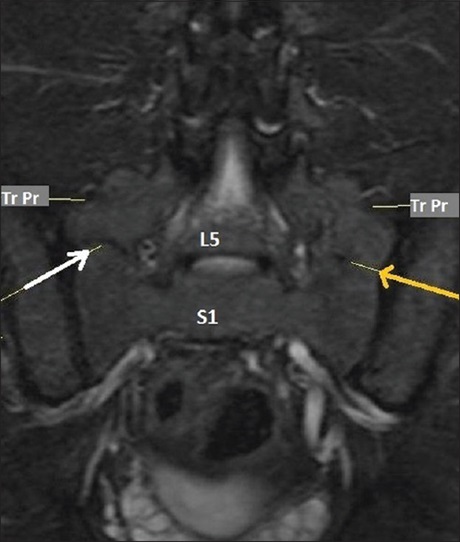
Materials and Methods: This prospective observational study includes 317 patients, referred for MRI of the lumbosacral spine. Vertebrae were counted manually using sagittal whole spine localizer images (Mobi View). Short tau inversion recovery (STIR) coronal images were included for classification of lumbosacral transitional vertebrae (LSTV). Prevalence and types of LSTV and level of proximal RRA, aortic bifurcation, and conus termination were documented.
Results: About 25.5% of patients showed LSTV and 7.8% of patients showed variation in presacral vertebral count without LSTV. Castellvi Type IIIb LSTV was most prevalent followed by Type IIb. There was significant variation in the level of aortic bifurcation, RRA, and conus termination in patients with normal count and with LSTV.
Conclusions: Spinal and paraspinal structures, such as aortic bifurcation, RRA, and conus termination, cannot be considered as landmark for numbering of vertebrae. Counting of vertebrae manually from C2 is recommended for confident documentation of numerical variation in vertebrae. The inclusion of STIR coronal image helps in better identification and characterization of the LSTV.
Downloads
Article Details
Section

This work is licensed under a Creative Commons Attribution-NonCommercial 4.0 International License.
This is an open access journal, and articles are distributed under the terms of the Creative Commons Attribution-NonCommercial-ShareAlike 4.0 License, which allows others to remix, tweak, and build upon the work non-commercially, as long as appropriate credit is given and the new creations are licensed under the identical terms.
How to Cite
References
1. Weiss KL, Storrs JM, Banto RB. Automated spine survey iterative scan technique. Radiology 2006;239:255‑62.
2. Carrino JA, Campbell PD Jr, Lin DC, Morrison WB, Schweitzer ME, Flanders AE, et al. Effect of spinal segment variants on numbering vertebral levels at lumbar MR imaging. Radiology 2011;259:196‑202.
3. Bailey W, Carter RA. Anomalies of the spine: A correlation of anatomical, roentgenological, and clinical findings. Cal West Med 1938;49:46‑52.
4. Hahn PY, Strobel JJ, Hahn FJ. Verification of lumbosacral segments on MR images: Identification of transitional vertebrae. Radiology 1992; 182:580‑1.
5. Akbar JJ, Weiss KL, Saafir MA, Weiss JL. Rapid MRI detection of vertebral numeric variation. AJR Am J Roentgenol 2010;195:465‑6.
6. Uçar D, Uçar BY, Cosar Y, Emrem K, Gümüssuyu G, Mutlu S, et al. Retrospective cohort study of the prevalence of lumbosacral transitional vertebra in a wide and well‑represented population. Arthritis 2013;2013:461425.
7. Apazidis A, Ricart PA, Diefenbach CM, Spivak JM. The prevalence of transitional vertebrae in the lumbar spine. Spine J 2011;11:858‑62.
8. Sekharappa V, Amritanand R, Krishnan V, David KS. Lumbosacral transition vertebra: Prevalence and its significance. Asian Spine J 2014; 8:51‑8.
9. Wigh RE, Anthony HF Jr. Transitional lumbosacral discs. Probability of herniation. Spine (Phila Pa 1976) 1981;6:168‑71.
10. Nicholson AA, Roberts GM, Williams LA. The measured height of the lumbosacral disc in patients with and without transitional vertebrae. Br J Radiol 1988;61:454‑5.
11. O’Driscoll CM, Irwin A, Saifuddin A. Variations in morphology of the lumbosacral junction on sagittal MRI: Correlation with plain radiography. Skeletal Radiol 1996;25:225‑30.
12. Castellvi AE, Goldstein LA, Chan DP. Lumbosacral transitional vertebrae and their relationship with lumbar extradural defects. Spine (Phila Pa 1976) 1984;9:493‑5.
13. Lee CH, Park CM, Kim KA, Hong SJ, Seol HY, Kim BH, et al. Identification and prediction of transitional vertebrae on imaging studies: Anatomical significance of paraspinal structures. Clin Anat 2007;20:905‑14.
14. Hughes RJ, Saifuddin A. Numbering of lumbosacral transitional vertebrae on MRI: Role of the iliolumbar ligaments. AJR Am J Roentgenol 2006;187:W59‑65.
15. Lee CH, Seo BK, Choi YC, Shin HJ, Park JH, Jeon HJ, et al. Using MRI to evaluate anatomic significance of aortic bifurcation, right renal artery, and conus medullaris when locating lumbar vertebral segments. AJR Am J Roentgenol 2004;182:1295‑300.
16. Tokgoz N, Ucar M, Erdogan AB, Kilic K, Ozcan C. Are spinal or paraspinal anatomic markers helpful for vertebral numbering and diagnosing lumbosacral transitional vertebrae? Korean J Radiol 2014; 15:258‑66.
17. Peh WC, Siu TH, Chan JH. Determining the lumbar vertebral segments on magnetic resonance imaging. Spine (Phila Pa 1976) 1999; 24:1852‑5.
18. Konin GP, Walz DM. Lumbosacral transitional vertebrae: Classification, imaging findings, and clinical relevance. AJNR Am J Neuroradiol 2010;31:1778‑86.
19. French HD, Somasundaram AJ, Schaefer NR, Laherty RW. Lumbosacral transitional vertebrae and its prevalence in the Australian population. Global Spine J 2014;4:229‑32.
20. Nardo L, Alizai H, Virayavanich W, Liu F, Hernandez A, Lynch JA, et al. Lumbosacral transitional vertebrae: Association with low back pain. Radiology 2012;265:497‑503.
21. Healy JC, Borley NR, Mundy AR, Collins P, Wigley C. True pelvis, pelvic floor and perineum. In: Standring S, editor. Gray’s Anatomy: The Anatomical Basis of Clinical Practice. 39th ed. New York: Elsevier Churchill Livingstone; 2005. p. 1360.
22. Chithriki M, Jaibaji M, Steele RD. The anatomical relationship of the aortic bifurcation to the lumbar vertebrae: A MRI study. Surg Radiol Anat 2002;24:308‑12.
23. Appaji AC, Kulkarni R, Pai B. Level of bifurcation of aorta and iliocaval confluence and its clinical relevance. IOSR J Dent Med Sci 2014; 13:56‑60.
24. Prakash, Mokhasi V, Rajini T, Shashirekha M. The abdominal aorta and its branches: Anatomical variations and clinical implications. Folia Morphol (Warsz) 2011;70:282‑6.
25. Saifuddin A, Burnett SJ, White J. The variation of position of the conus medullaris in an adult population. A magnetic resonance imaging study. Spine (Phila Pa 1976) 1998;23:1452‑6.
26. Soleiman J, Demaerel P, Rocher S, Maes F, Marchal G. Magnetic resonance imaging study of the level of termination of the conus medullaris and the thecal sac: Influence of age and gender. Spine (Phila Pa 1976) 2005;30:1875‑80.