

Associations of ultrasound splenic size and clinico‑laboratory values in patients with Sickle Cell Anemia at University College Hospital, Ibadan, Nigeria
Article Sidebar
Views | PDF/EPUB Downloads:
346
/ 91
/ 46
Main Article Content
Abstract
Background/Aims: Sickle cell anemia also known as haemoglobin SS (HbSS) is a genetic disease arising from the replacement of glutamic acid with valine at position 6 of the beta hemoglobin chain. This vaso-occlusive disease affects most of the organs in the body with the spleen commonly affected resulting in recurrent infarction. This study aims to assess the relationship between the ultrasound splenic length (LS) with the steady state packed cell volume (PCV), frequency of blood transfusion, and anthropometric parameters (weight and height) among patients with sickle cell anemia.
Materials and Methods: This is an observational cross-sectional study with 128 consenting HbSS patients recruited. Sickle cell anemia (HbSS) patients with no crises/illness within the last 4 weeks prior to the study period were included in the study. Patient’s demographics, steady PCV and ultrasound findings of the spleen were documented into the study pro forma. Data were analyzed using the Statistical package for the Social Sciences software version 21. Mean, median, standard deviation, and Chi-square were used.
A P ≤ 0.05 was considered statistically significant and a confidence interval (CI) of 95%.
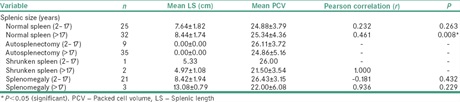
Results: The median age for all the patients was 19.00 years with a CI of 19.06–23.2 years. For children (2–17 years), the median age was 11.00 years with a CI of 9.35–11.36 years while for the adults was 28.00 years with a CI of 27.40–31.77 years. The median steady‑PCV obtained in this study was 25.0%. Adult HbSS patients with normal LS had a significant correlation with the steady PCV.
Conclusion: Normal-sized spleen on sonography may be a pointer to the steady PCV in sickle cell anemia patients. This study, therefore, suggests the need for sonography of the spleen in HbSS patients as part of their routine follow-up investigations.
Downloads
Article Details
Section
This is an open access journal, and articles are distributed under the terms of the Creative Commons Attribution-NonCommercial-ShareAlike 4.0 License, which allows others to remix, tweak, and build upon the work non-commercially, as long as appropriate credit is given and the new creations are licensed under the identical terms.
How to Cite
References
1. Al‑Salem AH. Splenic complications of sickle cell anemia and the role of splenectomy. ISRN Hematol 2011;2011:864257.
2. Gale HI, Bobbitt CA, Setty BN, Sprinz PG, Doros G, Williams DD, et al. Expected sonographic appearance of the spleen in children and
young adults with sickle cell disease: An Update. J Ultrasound Med 2016;35:1735‑45.
3. Facts | Sickle Cell Disease | NCBDDD | CDC [Internet]. Available from: https://www.cdc.gov/ncbddd/sicklecell/facts.html. [Last assessed on 2017 Oct 10].
4. Lane PA. Sickle cell disease. Pediatr Clin North Am 1996;43:639‑64.
5. World Health Organization. Sickle ‑cell Anaemia Report by the Secretariat Prevalence of Sickle Cell Anaemia; 2006. Available from:
http://apps.who.int/gb/archive/pdf_files/WHA59/A59_9‑en.pdf. [Last assessed on 2019 Sep 09].
6. Babadoko AA, Ibinaye PO, Hassan A, Yusuf R, Ijei IP, Aiyekomogbon J, et al. Autosplenectomy of sickle cell disease in Zaria, Nigeria: An ultrasonographic assessment. Oman Med J 2012;27:121‑3.
7. Aliyu ZY, Tumblin AR, Kato GJ. Current therapy of sickle cell disease. Haematologica 2006;91:7‑10.
8. Akingbola TS, Kolude B, Aneni EC, Raji AA, Iwara KU, Aken’Ova YA, et al. Abdominal pain in adult sickle cell disease patients: A Nigerian
experience. Ann Ib Postgrad Med 2011;9:100‑4.
9. Ojo O, Shokunbi W, Agunloye A. Splenic size in Sickle Cell Anaemia Patients in A Tertiary Hospital. Nigerian Hospital Practice. Niger Hosp
Pract 2007;13:82‑7.
10. Eze CU, Offordile GC, Agwuna KK, Ocheni S, Nwadike IU, Chukwu BF. Sonographic evaluation of the spleen among sickle cell disease patients in a teaching hospital in Nigeria. Afr Health Sci 2015;15:949‑58.
11. Attalla BI. Abdominal sonographic findings in children with sickle cell anemia. J Diagn Med Sonogr 2010;26:281‑5.
12. Thomas S, Sinha DN, Singh AK, Deopa D, Niranjan R. Morphometry of spleen in human fetuses at different gestational ages. Transl Res
Anat 2019;15:100042.
13. Chaudhry SR, Luskin V, Panuganti KK. Anatomy, Abdomen and Pelvis, Spleen. In: StatPearls. Treasure Island (FL): StatPearls Publishing; 2021.
14. Keith M, Arthur D. Abdomen. In: Glazer J, Odyniec C, editors. Clinically Oriented Anatomy. Lippincott Williams & Wilkin, Philadelphia: RR Donnelly and Sons Willard; 2006. p. 281‑4.
15. Brousse V, Buffet P, Rees D. The spleen and sickle cell disease: The sick (led) spleen. Br J Haematol 2014;166:165‑76.
16. Sickle Cell Disease: Practice Essentials, Background, Genetics . Available from: https://emedicine.medscape.com/article/205926‑
overview#a4 [Last accessed on 2022 Jun 23].
17. Luntsi G, Eze CU, Ahmadu MS, Bukar AA, Ochie K. Sonographic evaluation of some abdominal organs in sickle cell disease patients in a Tertiary health institution in Northeastern Nigeria. J Med Ultrasound 2018;26:31‑6.
18. West MS, Wethers D, Smith J, Steinberg M. Laboratory profile of sickle cell disease: A cross‑sectional analysis. The cooperative study of sickle cell disease. J Clin Epidemiol 1992;45:893‑909.
19. Akinbami A, Dosunmu A, Adediran A, Oshinaike O, Phillip A, Vincent O, et al. Steady state hemoglobin concentration and packed cell
volume in homozygous sickle cell disease patients in Lagos, Nigeria. Caspian J Intern Med 2012;3:405‑9.
20. Chou ST. Transfusion therapy for sickle cell disease: A balancing act. Hematology Am Soc Hematol Educ Program 2013;2013:439‑46.
21. Ehimwenma O, Tagbo MT. Determination of normal dimension of the spleen by ultrasound in an endemic tropical environment. Niger
Med J 2011;52:198‑203.
22. Spielmann AL, DeLong DM, Kliewer MA. Sonographic evaluation of spleen size in tall healthy athletes. AJR Am J Roentgenol 2005;184:45‑9.
23. Walczyk J, Walas MK. Errors made in the ultrasound diagnostics of the spleen. J Ultrason 2013;13:65‑72.
24. Wilson‑Okoh DA, Nwauche CA, Ejele OA. Splenic changes in sickle cell anaemia. Niger J Med 2006;15:20‑3.
25. Fasola FA, Adekanmi AJ. Haematological profile and blood transfusion pattern of patients with sickle cell anaemia vary with spleen size. Ann Ib Postgrad Med 2019;17:30‑8.
26. Brown BJ, Fatunde OJ, Sodeinde O. Correlates of steady‑state haematocrit and hepatosplenomegaly in children with sickle cell disease in Western Nigeria. West Afr J Med 2012;31:86‑91.
27. Yetunde A, Anyaegbu CC. Profile of the Nigerian sickle cell anaemia patients above 30 years of age. Cent Afr J Med 2001;47:108‑11.
28. Akinola NO, Bolarinwa RA, Faponle AF. The import of abdominal pain in adults with sickle cell disorder. West Afr J Med 2009;28:83‑6.
29. Adeodu OO, Adekile AD. Clinical and laboratory features associated with persistent gross splenomegaly in Nigerian children with sickle
cell anaemia. Acta Paediatr Scand 1990;79:686‑90.
30. Durosinmi MA, Salawu L, Ova YA, Lawal OO, Fadiran OA. Haematological parameters in sickle cell anaemia patients with and without splenomegaly. Niger Postgrad Med J 2005;12:271‑4.
31. Rosenberg HK, Markowitz RI, Kolberg H, Park C, Hubbard A, Bellah RD. Normal splenic size in infants and children: Sonographic measurements. AJR Am J Roentgenol 1991;157:119‑21.
32. Konuş OL, Ozdemir A, Akkaya A, Erbaş G, Celik H, Işik S. Normal liver, spleen, and kidney dimensions in neonates, infants, and children: Evaluation with sonography. AJR Am J Roentgenol 1998; 171: 1693‑8.