

Sonographic evaluation of the optic nerve sheath diameter and anterior chamber depth of the eye among apparently healthy adults in Kano, Nigeria
Article Sidebar
Views | PDF/EPUB Downloads:
431
/ 86
/ 38
Main Article Content
Abstract
Background: Measurement of optic nerve sheath diameter (ONSD) and anterior chamber depth (ACD) by ultrasound is increasingly used as a marker to detect raised intracranial pressure and other eye pathologies. Knowledge of normal ONSD and ACD in a healthy population is essential in the interpretation of pathological conditions.
Aim: The study aimed at evaluating the ONSD and ACD of the eye in apparently healthy adults in Kano State.
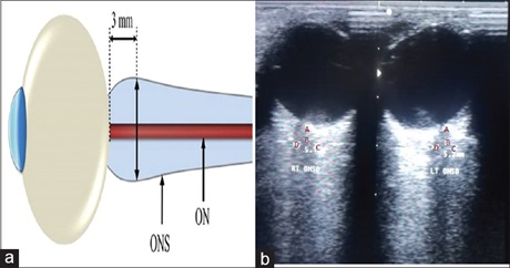
Materials and Methods: This was a prospective and cross-sectional study conducted among apparently healthy adults in Kano State from April 2019 to October 2019. Using convenience sampling method, 384 adults participated in the study. An ethical approval was obtained from the Human Research and Ethics Committee of the Kano State Ministry of Health, and informed consent was obtained from all the selected participants. A portable digital ultrasound machine, Nortek CS 3 with a 7.5 MHz linear transducer, was used to obtain ACD and ONSD at 3 mm behind the globe, and the values were recorded in data capture sheet. The obtained data were analyzed using SPSS version 23.0.
Results: The mean and standard deviation of the right and left ONSDs for males was 4.42 ± 1.38 mm and 4.44 ± 1.41 mm and for the females was 4.39 ± 1.31 mm and 4.41 ± 1.31, respectively. The mean and standard deviation of the right and left ACDs for males was 3.16 ± 0.37 mm and 3.14 ± 0.35 mm and for females was 3.12 ± 0.40 mm and 3.11 ± 1.39 mm, respectively.
Conclusion: The study has established normative values for the ONSD and ACD of the eye in Kano State, Nigeria.
Downloads
Article Details
Section
This is an open access journal, and articles are distributed under the terms of the Creative Commons Attribution-NonCommercial-ShareAlike 4.0 License, which allows others to remix, tweak, and build upon the work non-commercially, as long as appropriate credit is given and the new creations are licensed under the identical terms.
How to Cite
References
1. Kolade‑Yunusa HO, Itanyi U. Ultrasonograhic measurement of optic nerve sheath diameter in normal adults. Ann Int Med Den Res 2017;3:30‑4.
2. Shirodkar CG, Rao SM, Mutkule DP, Harde YR, Venkategowda PM, Mahesh MU. Optic nerve sheath diameter as a marker for evaluation and prognostication of intracranial pressure in Indian patients: An observational study. Indian J Crit Care Med 2014;18:728‑34.
3. Chun BY, Cestari DM. Advances in experimental optic nerve regeneration. Curr Opin Ophthalmol 2017;28:558‑63.
4. McCaa CS. The eye and visual nervous system: Anatomy, physiology and toxicology. Environ Health Perspect 1982;44:1‑8.
5. Hassen GW, Sweeney B, Portillo T, Ali D, Nazeer O, Habal R, et al. Anterior chamber depth measurement using ultrasound to assess elevated intraocular pressure. Am J Emerg Med 2015;33:860.e1‑3.
6. Chen LM, Wang LJ, Hu Y, Jiang XH, Wang YZ, Xing YQ. Ultrasonic measurement of optic nerve sheath diameter: A non‑invasive surrogate
approach for dynamic, real‑time evaluation of intracranial pressure. Br J Ophthalmol 2019;103:437‑41.
7. Toscano M, Spadetta G, Pulitano P, Rocco M, Di Piero V, Mecarelli O, et al. Optic nerve sheath diameter ultrasound evaluation in intensive care
unit: Possible role and clinical aspects in neurological critical patients’ daily monitoring. Biomed Res Int 7;2017:1‑7.
8. Murphy DL, Oberfoell SH, Trent SA, French AJ, Kim DJ, Richards DB. Validation of a low‑cost optic nerve sheath ultrasound phantom: An
educational tool. J Med Ultrasound 2017;25:96‑100.
9. Özmen Z, Aktaş F, Özmen ZC, Almus E, Demir O. Ultrasound measurement of liver longitudinal length in a North Anatolian population: A community‑based study. Niger J Clin Pract 2018;21:653‑7.
10. Nelson E, Mulugeta L, Myers J. Microgravity‑induced fluid shift and ophthalmic changes. Life 2014;4:621‑65.
11. Shen M, Wang MR, Yuan Y, Chen F, Karp CL, Yoo SH, et al. SD‑OCT with prolonged scan depth for imaging the anterior segment of the eye. Ophthalmic Surg Lasers Imaging 2010;41 Suppl: S65‑9.
12. Zong Y, Xu Q, Jiang C, Zhu H, Yu J, Sun X. Measurement of and factors associated with the anterior chamber volume in healthy Chinese
adults. J Ophthalmol 2017;2017: 1‑6.
13. Maude RR, Amir HM, Hassan MU, Osbourne S, Sayeed KL, Karim MR. Trans‑orbital sonographic evaluation of normal optic nerve sheath diameter in healthy volunteers in Bangladesh. PLoS One 2013;8:e81013.
14. Chan PY, Mok KL. Transortital sonographic evaluation of optic nerve sheath diameter in normal Hong Kong Chinese adults. Hong Kong J
Emerg Med 2008;2008:197‑204.
15. Asghar A, Hashmi M, Hussain A. Optic nerve sheath diameter evaluated by transorbital sonography in healthy volunteers from Pakistan. Anaesthesia Pain Intensive Care 2015;19:282‑6.
16. Sedaghat MR, Mohammad Zadeh V, Fadakar K, Kadivar S, Abrishami M. Normative values and contralateral comparison of anterior chamber parameters measured by Pentacam and its correlation with corneal biomechanical factors. Saudi J Ophthalmol 2017;31:7‑10