

Sonographic estimation of gestational age using transverse cerebellar diameter among late trimester pregnancies in Kano, Northwest Nigeria
Article Sidebar
Views | PDF/EPUB Downloads:
120
/ 24
/ 10
Main Article Content
Abstract
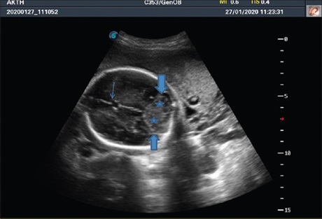
Background/Context: Accurate estimation of fetal gestational age (GA) is of paramount importance in the management of all pregnancies, especially for the planning of mode of the delivery and management of high-risk pregnancies. Obstetric ultrasonography is a simple, available, affordable, and noninvasive modality for estimating fetal GA due to its high safety profile. The fetal cerebellum is easily visualized and reliably identified on ultrasound in the posterior cranial fossa after 14 weeks of gestation.
Aim: The aim of this study was to evaluate the utility of transverse cerebellar diameter (TCD) in determining GA in second- and third-trimester pregnancies and compare its accuracy with other established sonographically derived fetal biometric parameters.
Materials and Methods: A total of 424 pregnant women in their second and third trimesters who were sure of their last menstrual period (LMP) were recruited into the study over a period of 6 months (May to November 2019) at the Radiology Department of AKTH-Kano, Nigeria. The corresponding fetal TCD and other established fetal biometric indices (biparietal diameter [BPD], head circumference [HC], abdominal circumference [AC], and femur length [FL]) were sonographically obtained and correlated. Pearson’s bivariate coefficient was used to establish the correlation between the traditional biometric indices with TCD and GA derived by LMP. Multivariate linear regression analysis was used to assess the accuracy of the studied indices in predicting GA.
Results: The range for TCD in second- and third-trimester fetuses was 15.9–57.5 mm. The TCD parameter was more accurate (±1.753 days) than BPD (±2.298 days), HC (±2.337 days), and AC (±4.342 days) and marginally less accurate than FL (±1.165 days) in predicting GA among study subjects (P < 0.001).
Conclusion: TCD is a reliable and accurate parameter for GA estimation in late second- and third-trimester pregnancies when compared with established fetal biometric parameters among pregnant women in Kano, Nigeria.
Downloads
Article Details
Section
This is an open access journal, and articles are distributed under the terms of the Creative Commons Attribution-NonCommercial-ShareAlike 4.0 License, which allows others to remix, tweak, and build upon the work non-commercially, as long as appropriate credit is given and the new creations are licensed under the identical terms.
How to Cite
References
1. Ronald MR, editor. Blog: Evaluation of Gestation: Overview, Clinical Methods of Estimating Gestational Age. Virginia: Medscape Publishing Group; 2021. Available from: https://www.emedicine.medscape.com/article/259269‑overview?form=fpf. [Last accessed on 2022 Feb 24].
2. The Global Library of Women’s Medicine [Internet]. Carlisle (UK): International federation of Gynecology and Obstetrics, Inc.; Assessment
of Gestational Age by Ultrasound; c2008. Available from: https://www.glowm.com/section‑view/heading/Assessment%20of%20Gestational%20Age%20by%20Ultrasound/item/206. [Last accessed on 2022 Feb 23].
3. Shan BP, Madheswaran M. Revised estimates of ultrasonographic markers for gestational age assessment of singleton pregnancies among Indian population. Int J Adv Sci Technol 2010;17:1‑12.
4. Dietrich CF, Bolondi L, Duck F, Evans DH, Ewertsen C, Fraser AG, et al. History of ultrasound in medicine from its birth to date (2022), on occasion of the 50 years anniversary of EFSUMB. A publication of the European Federation of Societies for Ultrasound in Medicine and Biology (EFSUMB), designed to record the historical development of medical ultrasound. Med Ultrason 2022;24:434‑50.
5. Houston L, Newman R. Fetal ultrasound: How to put safety first. Contemp Obstet Gynaecol 2011;56:36‑43. Available from: https:// www.researchgate.net/publication/288063552_Fetal_ultrasound_How_to_put_safety_first. [Last accessed on 2022 Mar 03].
6. Eze CU, Onwuzu QE, Nwadike IU. Sonographic reference values for fetal transverse cerebellar diameter in the second and third trimesters in a Nigerian population. J Diagn Med Sonogr 2017;33:174‑81.
7. Bansal M, Bansal A, Jain S, Khare S, Ghai R. A study of correlation of transverse cerebellar diameter with gestational age in the normal & growth restricted fetuses in Western Uttar Pradesh. People’s J Sci Res 2014;7:2‑7.
8. Miller DL. Safety assurance in obstetrical ultrasound. Semin Ultrasound CT MR 2008;29:156‑64.
9. Houston LE, Odibo AO, Macones GA. The safety of obstetrical ultrasound: A review. Prenat Diagn 2009;29:1204‑12.
10. Lerner JP. Fetal growth and well‑being. Obstet Gynecol Clin North Am 2004;31:159‑76.
11. Adeyekun AA, Orji MO. Predictive accuracy of transcerebellar diameter in comparison with other foetal biometric parameters for
gestational age estimation among pregnant Nigerian women. East Afr Med J 2014;91:138‑44.
12. Shivalingaiah N, Nirmala K, Sowmya R, Ananya T, Kanmani P, Marimuthu TF, et al. Kidney length as a parameter for determination of gestational age in pregnancy. Int J Reprod Contracept Obstet Gynecol 2014;3:424.
13. Goel P, Singla M, Ghai R, Jain S, Budhiraja V, Ramesh CS. Transverse cerebellar diameter – A marker for estimation of gestational age. J Anat Soc India 2010;59:158‑61.
14. Sharma G, Ghode R. Fetal transcerebellar diameter and transcerebellar diameter – Abdominal circumference ratio as a menstrual age independent parameter for gestational age estimation with grading of cerebellar maturity. Int J Reprod Contracept Obstet Gynecol 2015; 4: 2036‑40.
15. Prasad VN, Dhakal V, Chhetri PK. Accuracy of transverse cerebellar diameter by ultrasonography in the evaluation gestational age of fetus. Int J Adv Med 2017;4:836‑41.
16. Utoo BT, Farouk HU, Muhammed RL, Bako B, Azeez OA, Joshua TG, et al. Unsure last normal menstrual period among pregnant women in low resource setting: The experience from federal teaching hospital Gombe, North‑Eastern Nigeria. J Women Health Care Issues 2022; 5: 133‑41. Available from: https://www.auctoresonline.org/article/unsure‑last‑normal‑menstrual‑period‑among‑pregnant‑women‑in‑low‑resource‑setting‑the‑experience‑from‑federal‑teaching‑hospital‑gombe‑north‑eastern‑nigeria. [Last accessed on 2022 Dec 16]. [doi: 10.31579/2642‑9756/133].
17. Hazra A, Gogtay N. Biostatistics series module 5: Determining sample size. Indian J Dermatol 2016;61:496‑504.
18. Ravindernath ML, Reddy M, Reddy N. Accuracy of transverse cerebellar diameter measurement by ultrasonography in the evaluation
of fetal age. Int J Adv Med 2017;4:836‑41.
19. Pavithra SN, Vimala D, Prema G, Shankar R. Determination of gestational age: Correlation between fetal biometry and transverse cerebellar diameter in women with uncomplicated pregnancy. Inter J Reprod Contracept Obstet Gynecol 2017;6:3599‑608. Available:
https://link.gale.com/apps/doc/A534838741/HRCA?u=anon~b0a1ef07&sid=googleScholar&xid=4019a9c9. [Last accessed on 2022 Mar 28].
20. Agrawal C, Agrawal KK, Gandhi S, Chaudhary S. Correlation between ultrasonography measured transcerebellar diameter of fetus with early and late gestational age. Int J Reprod Contracept Obstet Gynecol 2015;4:2010‑3.
21. Sumanta KM, Sandip KG, Saikat R, Barun P. Evaluation of fetal transcerebellar diameter as a sonological parameter for the estimation of fetal gestational age in comparison to biparietal diameter and femur length. IAIM 2019;6:41‑50.
22. Mahmoud MZ, Mahmoud OA, Abdulla AA. Fetal transverse cerebellar diameter measurement for prediction of gestational age in pregnant Sudanese ladies. Int J Life Sci Med Res 2013;3:89‑93.
23. Fagbamigbe AF, Bamgboye EA, Yusuf BO, Akinyemi JO, Issa BK, Ngige E, et al. The Nigeria wealth distribution and health seeking behaviour: Evidence from the 2012 National HIV/AIDS and reproductive health survey. Health Econ Rev 2015;5:5.
24. Naseem F, Fatima N, Yasmeen S, Saleem S. Comparison between transcerebellar diameter with biparietal diameter of ultrasound for gestational age measurement in third trimester of pregnancy. J Coll Physicians Surg Pak 2013;23:322‑5.
25. Nagesh R, Seetha PV, Shukla AK. Transverse cerebellar diameter – An ultrasonographic parameter for estimation of fetal gestational age. Int J Contemp Med Res 2016;3:1029‑31.