

Pattern of prenatal ultrasound diagnosed anterior abdominal wall defects at the University College Hospital, Ibadan, Nigeria: A pictorial essay
Article Sidebar
Views | PDF/EPUB Downloads:
380
/ 87
/ 40
Main Article Content
Abstract
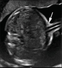
Anterior abdominal wall defects form a wide spectrum of congenital abnormalities that allow the abdominal contents to protrude through an unusual opening on the abdominal wall. These defects could be physiological or pathological depending on the time of diagnosis. They include physiological gut herniation, congenital umbilical cord hernia, omphalocele, gastroschisis, ectopia cordis, bladder exstrophy, body-stalk anomaly, Prune-Belly Syndrome, and pentalogy of Cantrell. Correct prenatal diagnosis of these anomalies with ultrasound (US) is extremely important for patient management. Evaluation of the defect relative to the umbilical cord insertion site is fundamentally important in differentiating among the various malformations. We present a pictorial essay of the spectrum of anterior abdominal wall defects diagnosed prenatally with US seen over a 5-year period at the University
College Hospital, Ibadan.
Downloads
Article Details
Section
This is an open access journal, and articles are distributed under the terms of the Creative Commons Attribution-NonCommercial-ShareAlike 4.0 License, which allows others to remix, tweak, and build upon the work non-commercially, as long as appropriate credit is given and the new creations are licensed under the identical terms.
How to Cite
References
1. Pakdaman R, Woodward PJ, Kennedy A. Complex abdominal wall defects: Appearances at prenatal imaging. Radiographics 2015;35:636‑49.
2. Emanuel PG, Garcia GI, Angtuaco TL. Prenatal detection of anterior abdominal wall defects with US. Radiographics 1995;15:517‑30.
3. Sadler TW. The embryologic origin of ventral body wall defects. Semin Pediatr Surg 2010;19:209‑14.
4. American Institute of Ultrasound in Medicine. AIUM practice guideline for the performance of obstetric ultrasound examinations. J Ultrasound Med 2013;32:1083‑101.
5. Salomon LJ, Alfirevic Z, Berghella V, Bilardo C, Hernandez‑Andrade E, Johnsen SL, et al. Practice guidelines for performance of the routine mid‑trimester fetal ultrasound scan. Ultrasound Obstet Gynecol 2011;37:116‑26.
6. Moore KL, Persaud TV, Torchia MG. Fourth to eighth weeks of human development. In: The Developing Human: Clinically Oriented Embryology. 9th ed. Philadelphia, Pa: Saunders; 2013.
7. Moore KL, Persaud TV, Torchia MG. Alimentary system. In: The Developing Human: Clinically Oriented Embryology. 9th ed. Philadelphia, Pa: Saunders; 2013.
8. Richards DS, Kays DW. Prenatal ultrasonographic diagnosis of a simple umbilical hernia. J Ultrasound Med 1998;17:265‑7.
9. Evans AG. The comparative incidence of umbilical hernias in colored and white infants. J Natl Med Assoc 1941;33:158‑60.
10. Jackson OJ, Moglen LH. Umbilical hernia. A retrospective study. Calif Med 1970;113:8‑11.
11. Cohen‑Overbeek TE, Tong WH, Hatzmann TR, Wilms JF, Govaerts LC, Galjaard RJ, et al. Omphalocele: Comparison of outcome following prenatal or postnatal diagnosis. Ultrasound Obstet Gynecol 2010;36:687‑92.
12. Salihu HM, Boos R, Schmidt W. Omphalocele and gastrochisis. J Obstet Gynaecol 2002;22:489‑92.
13. Grigore M, Iliev G, Gafiteanu D, Cojocaru C. The fetal abdominal wall defects using 2D and 3D ultrasound. Pictorial essay. Med Ultrason 2012;14:341‑7.
14. Stoll C, Alembik Y, Dott B, Roth MP. Omphalocele and gastroschisis and associated malformations. Am J Med Genet A 2008;146A:1280‑5.
15. Barisic I, Clementi M, Häusler M, Gjergja R, Kern J, Stoll C, et al. Evaluation of prenatal ultrasound diagnosis of fetal abdominal wall defects by 19 European registries. Ultrasound Obstet Gynecol 2001;18:309‑16.
16. Snyder CL. Outcome analysis for gastroschisis. J Pediatr Surg 1999;34:1253‑6.
17. Cerekja A, Piazze J, Cozzi D. Early prenatal sonographic diagnosis of gastroschisis. J Clin Ultrasound 2012;40:526‑8.
18. Ibadin MO, Ademola AA, Ofovwe GE. Familial prune Belly syndrome in a Nigerian family. Saudi J Kidney Dis Transpl 2012;23:338‑42.
19. Okeniyi JA, Ogunlesi TA, Dedeke IO, Oyelami OA, Oyedeji GA. Prune Belly syndrome in a Nigeria child. Internet J Pediatr Neonatol 2005;5:2.
20. Agarwal R. Prenatal diagnosis of abdominal wall defects. Indian J Radiol Imaging 2005;15:361‑72.
21. Zugor V, Schott GE, Labanaris AP. The Prune Belly syndrome: Urological aspects and long‑term outcomes of a rare disease. Pediatr Rep 2012;4:e20.
22. Bugge M. Body stalk anomaly in Denmark during 20 years (1970‑1989). Am J Med Genet A 2012;158A:1702‑8.
23. Russo R, D’Armiento M, Angrisani P, Vecchione R. Limb body wall complex: A critical review and a nosological proposal. Am J Med Genet 1993;47:893‑900.
24. Lockwood CJ, Scioscia AL, Hobbins JC. Congenital absence of the umbilical cord resulting from maldevelopment of embryonic body folding. Am J Obstet Gynecol 1986;155:1049‑51.
25. Daskalakis G, Sebire NJ, Jurkovic D, Snijders RJ, Nicolaides KH. Body stalk anomaly at 10‑14 weeks of gestation. Ultrasound Obstet Gynecol 1997;10:416‑8.
26. Shah AK, Joshi MA, Kumar S. Bladder exstrophy – A case report. Indian J Radiol Imaging 2006;16:103‑6.
27. Siffel C, Correa A, Amar E, Bakker MK, Bermejo‑Sánchez E, Bianca S, et al. Bladder exstrophy: An epidemiologic study from the international clearinghouse for birth defects surveillance and research, and an overview of the literature. Am J Med Genet C Semin Med Genet 2011;157C:321‑32.
28. Lee EH, Shim JY. New sonographic finding for the prenatal diagnosis of bladder exstrophy: A case report. Ultrasound Obstet Gynecol 2003;21:498‑500.
29. Gearhart JP, Ben‑Chaim J, Jeffs RD, Sanders RC. Criteria for the prenatal diagnosis of classic bladder exstrophy. Obstet Gynecol 1995;85:961‑4.