

Ultrasound reference values for Inferior Vena Cava diameter and Collapsibility Index among adult Nigerians
Article Sidebar
Views | PDF/EPUB Downloads:
332
/ 94
/ 46
Main Article Content
Abstract
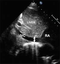
Background: Correct estimation of intravascular volume is crucial in critically ill and traumatized patients. Measurement of the central venous pressure (CVP) is invasive and time consuming. Studies have shown that inferior vena cava diameter (IVCD) correlates with CVP. Sonographic assessment of IVCD and its respirophasic changes (collapsibility index; CI) is a non-invasive, quick and reliable means of estimating CVP and hence, intravascular fluid volume. Data on such studies are scanty among adult Nigerians.
Aim: To establish normograms of IVCD and CI for healthy adults in Benin City, Nigeria as well as determine the relationship of IVCD and CI with height, weight, body mass index (BMI), age and gender.
Method: Four hundred apparently healthy adult volunteers were prospectively studied by means of ultrasound. Demographic data and BMI were obtained. The IVCD was measured during inspiration, expiration and sniff. The CI was subsequently calculated for each subject. Statistical Package for the Social Sciences (SPSS) version 17.0 was used for data analysis including tests of significance. Probability values less than or equal to 0.05 were considered significant.
Results: The mean IVCD in this study was 6.1±2.2mm and 13.0±4.0 mm for inspiration and expiration respectively. The mean CI was 49.7±0.5%. There was no statistically significant correlation between IVCD and CI with height and BMI.
Conclusion: This study has determined normal IVCD and CI reference range for healthy Nigerian adults. The CI is independent of height, weight, BMI and gender. Since the CI is not dependent on physical attributes and gender, it may serve as an objective tool for monitoring the fluid status of patient
Downloads
Article Details
Section
This is an open access journal, and articles are distributed under the terms of the Creative Commons Attribution-NonCommercial-ShareAlike 4.0 License, which allows others to remix, tweak, and build upon the work non-commercially, as long as appropriate credit is given and the new creations are licensed under the identical terms.
How to Cite
References
1. Kastrup M, Markewitz A, Spies C, Carl M, Erb J, Grosse J, et al. Current practice of hemodynamic monitoring and vasopressor and inotropic therapy in post‑operative cardiac surgery patients in Germany: Results from a postal survey. Acta Anaesthesiol Scand 2007;51:347‑58.
2. McIntyre LA, Hébert PC, Fergusson D, Cook DJ, Aziz A, Canadian Critical Care Trials Group, et al. A survey of Canadian intensivists’ resuscitation practices in early septic shock. Crit Care 2007;11:R74.
3. David R, Adam F. Clinical Medicine: A Clerking Companion. New York: Oxford University Press; 2011. p. 134‑5.
4. Mark JA. Ultrasound measurement of the inferior vena cava can predict shock. J Trauma 2007;63:124‑5.
5. Field JM, Braster MJ. The Textbook of Emergency Cardiovascular Care and CPR. Philadelphia: Lippincott William and Wilkins; 2009. p. 130‑2.
6. Lyon M, Blaivas M, Brannam L. Sonographic measurement of the inferior vena cava as a marker of blood loss. Am J Emerg Med 2005;23:45‑50.
7. Natori H, Tamaki S, Kira S. Ultrasonographic evaluation of ventilatory effect on inferior vena caval configuration. Am Rev Respir Dis 1979;120:421‑7.
8. Simonson JS, Schiller NB. Sonospirometry: A new method for noninvasive estimation of mean right atrial pressure based on two‑dimensional echographic measurements of the inferior vena cava during measured inspiration. J Am Coll Cardiol 1988;11:557‑64.
9. Mintz GS, Kotler MN, Parry WR, Iskandrian AS, Kane SA. Real‑time inferior vena caval ultrasonography: Normal and abnormal findings and its use in assessing right‑heart function. Circulation 1981;64:1018‑25.
10. Moreno FL, Hagan AD, Holmen JR, Pryor TA, Strickland RD, CastleCH, et al. Evaluation of size and dynamics of the inferior vena cava as an index of right‑sided cardiac function. Am J Cardiol 1984;53:579‑85.
11. Kircher BJ, Himelman RB, Schiller NB. Noninvasive estimation of right atrial pressure from the inspiratory collapse of the inferior vena cava. Am J Cardiol 1990;66:493‑6.
12. Pinsky MR, Brochurd L, Mancebo. Applied Physiology in Intensive Care Medicine. 2nd ed. Berlin: Springer Verlag; 2009. p. 181‑6.
13. American College of Emergency Physicians. Emergency ultrasound guidelines. Ann Emerg Med 2009;53:550‑70.
14. Rudski LG, Lai WW, Afilalo J, Hua L, Handschumacher MD, Chandrasekaran K, et al. Guidelines for the echocardiographic assessment of the right heart in adults: A report from the American Society of Echocardiography endorsed by the European Association of Echocardiography, a registered branch of the European Society of Cardiology, and the Canadian Society of Echocardiography. J Am Soc Echocardiogr 2010;23:685‑713.
15. Mandelbaum A, Ritz E. Vena cava diameter measurement for estimation of dry weight in haemodialysis patients. Nephrol Dial Transplant 1996;11 Suppl 2:24‑7.
16. Grant E, Rendano F, Sevinc E, Gammelgaard J, Holm HH, Grønvall S, et al. Normal inferior vena cava: Caliber changes observed by dynamic ultrasound. AJR Am J Roentgenol 1980;135:335‑8.
17. Yanagawa Y, Sakamoto T, Okada Y. Hypovolemic shock evaluated by sonographic measurement of the inferior vena cava during resuscitation in trauma patients. J Trauma 2007;63:1245‑8.
18. Sefidbakht S, Assadsangabi R, Abbasi HR, Nabavizadeh A. Sonographic measurement of the inferior vena cava as a predictor of shock in trauma patients. Emerg Radiol 2007;14:181‑5.
19. Blehar DJ, Dickman E, Gaspari R. Identification of congestive heart failure via respiratory variation of inferior vena cava diameter. Am J Emerg Med 2009;27:71‑5.
20. Feissel M, Michard F, Faller JP, Teboul JL. The respiratory variation in inferior vena cava diameter as a guide to fluid therapy. Intensive Care Med 2004;30:1834‑7.
21. Dambatta AH. B‑mode ultrasonographic measurement of inferior vena cava among healthy adults in Kano, Nigeria. Niger J Basic Clin Sci 2016;13:94‑8.
22. Glockner JF, Lee CU. Magnetic resonance venography. Appl Radiol J 2010;39:6‑10.