

Computed tomographic findings of the brain in adult HIV‑infected patients at Doctor George Mukhari Academic Hospital, Ga‑Rankuwa, Pretoria, South Africa
Article Sidebar
Views | PDF/EPUB Downloads:
377
/ 115
/ 47
Main Article Content
Abstract
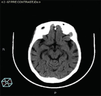
Background/Aim: The aim of this study is to determine the pattern of computed tomographic (CT) findings in HIV-infected patients referred for CT brain at Doctor George Mukhari Academic Hospital (DGMAH) and to correlate the CD4 counts with CT brain findings of the patients on antiretroviral (ARV) drugs, and those that are ARV naïve.
Methods: A descriptive, retrospective review of CT brains obtained from 128 slices Philips and GE, CT scanners, medical records, and laboratory results of 364 adult HIV-infected patients over a 6-month period (October 1, 2016–March 31, 2017) was conducted at Radiology Department of DGMAH. Statistical analyses were made using a Statistical Program for Social Sciences software (SPSS version 19.0).
Results: From the 364 CT brain findings of HIV-infected patients reviewed, 46.2% were male and 53.8% were female. The findings were as follows: brain atrophy (168; 46.2%); infarcts (55; 15.1%); hydrocephalus (24; 6.6%); white matter disease (18; 4.9%); mass lesions (13; 3.6%); rim enhancing lesions (12; 3.3%); intracranial bleed (11; 3.0%); tuberculous granuloma (32; 8.8%); tuberculous meningitis (15; 4.1%); and cryptococcal meningitis (2; 0.5%). Opportunistic infections and mass lesions still predominate at CD4
count <200 cells/mm3 although the reduction in the prevalence of opportunistic infections was observed. Brain infarct was seen at CD4 count <200 cells/mm3, and brain atrophy was seen at all CD4 count levels (median= 84 cells/mm3).
Conclusion: This study was conducted in the post-highly active ARV therapy era, and the most common CT scan brain finding was brain atrophy, followed by brain infarct.
Downloads
Article Details
Section
This is an open access journal, and articles are distributed under the terms of the Creative Commons Attribution-NonCommercial-ShareAlike 4.0 License, which allows others to remix, tweak, and build upon the work non-commercially, as long as appropriate credit is given and the new creations are licensed under the identical terms.
How to Cite
References
1. de Almeida SM, Letendre S, Ellis R. Human immunodeficiency virus and the central nervous system. Braz J Infect Dis 2006;10:41‑50.
2. Morley N. UNAIDS/WHO – Reports & Facts Sheet; 13 June, 2016. Available from: http://www.unaids.org/en/resources/fact‑sheet. [Last
accessed on 2017 Apr 18].
3. Sibtain NA, Chinn RJ. Imaging of the central nervous system in HIV infection. Imaging 2002;14:48‑59.
4. Graham CB 3rd, Wippold FJ 2nd, Pilgram TK, Fisher EJ, Smoker WR. Screening CT of the brain determined by CD4 count in HIV‑positive
patients presenting with headache. AJNR Am J Neuroradiol 2000;21:451‑4.
5. Hongsakul K, Laothamatas J. Computer tomographic findings of the brain in HIV‑patients at Ramathibodi hospital. J Med Assoc Thai
2008;91:895‑907.
6. Eze KC, Eze EU. Brain computed tomography of patients with HIV/AIDS before the advent of subsidized treatment program in
Nigeria. Niger Med J 2012;53:231‑5.
7. Bhigjee AI, Naidoo K, Patel VB, Govender D. Intracranial mass lesions in HIV‑positive patients – The kwaZulu/Natal experience.
Neuroscience AIDS research group. S Afr Med J 1999;89:1284‑8.
8. Dean D, Berger JR. Neuro‑AIDS in the developing world. Neurology2012;78:499‑500.
9. UNAIDS. South Africa – HIV and AIDS Estimates (South Africa); 2015. Available from: http://www.unaids.org/en/regionscountries/
countries/southafrica. [Last accessed on 2017 Apr 15].
10. Asselman V, Thienemann F, Pepper DJ, Boulle A, Wilkinson RJ, Meintjes G, et al. Central nervous system disorders after starting
antiretroviral therapy in South Africa. AIDS 2010;24:2871‑6.
11. Rothman RE, Keyl PM, McArthur JC, Beauchamp NJ Jr., Danyluk T, Kelen GD. A decision guideline for emergency department utilization
of noncontrast head computed tomography in HIV‑infected patients. Acad Emerg Med 1999;6:1010‑9.
12. Senocak E, Oğuz KK, Ozgen B, Kurne A, Ozkaya G, Unal S, et al. Imaging features of CNS involvement in AIDS. Diagn Interv Radiol
2010;16:193‑200.
13. Srivastan S. HIV Infection Increases Risk of Brain Shrinkage. “The Aids Beacon” – An Independent, Up‑to‑Date News and Information about HIV and AIDS, New Jersey, USA; 29 February, 2012. Available from: http://www.aidsbeacon.com. [Last accessed on2018 Jan 10].
14. World Health Organization. Global Tuberculosis Report 2014. Geneva: WHO; 2014: Available from: http://www.who.int/tb/publications/
global_report/gtbr14_main_text.pdf. [Last accessed on 2017 Sept 23].
15. Singer EJ, Valdes‑Sueiras M, Commins DL, Yong W, Carlson M. HIV stroke risk: Evidence and implications. Ther Adv Chronic Dis 2013;4:61‑70.
16. Mlay M, Bakari M. The prevalence of HIV among patients admitted with stroke at the Muhimbili National Hospital, Dares Salaam, Tanzania. Tanzan J Health Res 2010;12:1‑12.
17. Ortiz G, Koch S, Romano JG, Forteza AM, Rabinstein AA. Mechanisms of ischemic stroke in HIV‑infected patients. Neurology 2007;68:1257‑61.
18. Cole JW, Pinto AN, Hebel JR, Buchholz DW, Earley CJ, Johnson CJ, et al. Acquired immunodeficiency syndrome and the risk of stroke.
Stroke 2004;35:51‑6.
19. Hsue PY, Lo JC, Franklin A, Bolger AF, Martin JN, Deeks SG, et al. Progression of atherosclerosis as assessed by carotid intima‑media
thickness in patients with HIV infection. Circulation 2004;109:1603‑8.