

Head Computed Tomography: Dose Output and Relationship with Anthropotechnical Parameters
Article Sidebar
Views | PDF/EPUB Downloads:
77
/ 7
/ 8
Main Article Content
Abstract
Background: The number of computed tomography (CT) centers and examinations in Nigeria has shown a steady increase. This will
increase the collective dose and may potentially result in an increased incidence of cancer, hereditary diseases, and the possibility
of mild deterministic effects.
Objective: To determine radiation dose output and its relationship with anthropotechnical parameters.
Methodology: A retrospective analyses of digital CT files. Effective dose was derived from the dose‑length product and factor for
examination of head CT (0.0023 mSv/mGy‑cm). SPSS version 20.0 (SPSS Inc., Chicago, IL, USA) was used to analyze the data.
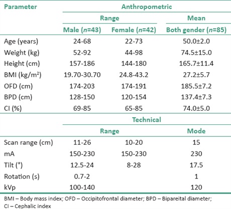
Results: Files of 43 male and 42 female (n = 85) adult patients were analyzed. The mean (and 75th percentile) of the CT dose index (CTDI), dose‑length product (DLP), and effective dose in noncontrast examinations were 48 (59) mGy, 874 (1301) mGy‑cm, and 1.8 (2.7) mSv, respectively. Contrast examinations yielded 54 (61) mGy, 1476 (2044) mGy‑cm, and 3.1 (4.3) mSv, respectively. DLP showed a weak relationship with BPD (r = −0.220), age (r = 0.211), cephalic index (r = −0.186), height (r = 0.158), and gantry tilt (r = 0.154). There was no relationship with weight (r = 0.076), range (r = −0.073), occipitofrontal diameter (r = 0.037), and body mass index (r = −0.018). The correlations were neither statistically nor clinically significant.
Conclusion: The CTDI is comparable with local values while the DLP is lower by a range of 5–31% but higher than foreign values by a range of 19–35%. Further optimization of CT radiation dose should be explored to eliminate the gulf between local and foreign dose outputs.
Downloads
Article Details
Section

This work is licensed under a Creative Commons Attribution-NonCommercial 4.0 International License.
This is an open access journal, and articles are distributed under the terms of the Creative Commons Attribution-NonCommercial-ShareAlike 4.0 License, which allows others to remix, tweak, and build upon the work non-commercially, as long as appropriate credit is given and the new creations are licensed under the identical terms.
How to Cite
References
1. van der Molen AJ, Schilham A, Stoop P, Prokop M, Geleijns J. A national survey on radiation dose in CT in the Netherlands. Insights Imaging 2013;4:383‑90.
2. Mettler FA Jr., Huda W, Yoshizumi TT, Mahesh M. Effective doses in radiology and diagnostic nuclear medicine: A catalog. Radiology 2008; 248:254‑63.
3. Amis ES Jr., Butler PF, Applegate KE, Birnbaum SB, Brateman LF, Hevezi JM, et al. American College of Radiology white paper on radiation dose in medicine. J Am Coll Radiol 2007;4:272‑84.
4. National Council on Radiation Protection and Measurements. Ionizing Radiation Exposure of the Population of the United States, NCRP Report No. 93. Bethesda, MD: National Council on Radiation Protection and Measurements; 1987.
5. Brenner DJ, Hall EJ. Computed tomography – An increasing source of radiation exposure. N Engl J Med 2007;357:2277‑84.
6. McCollough CH, Primak AN, Braun N, Kofler J, Yu L, Christner J. Strategies for reducing radiation dose in CT. Radiol Clin North Am 2009; 47:27‑40.
7. Vassileva J, Rehani MM, Al‑Dhuhli H, Al‑Naemi HM, Al‑Suwaidi JS, Appelgate K, et al. IAEA survey of pediatric CT practice in 40 countries in Asia, Europe, Latin America, and Africa: Part 1, frequency and appropriateness. AJR Am J Roentgenol 2012;198:1021‑31.
8. Garba I, Engel‑Hills P, Davidson F, Tabari AM. Computed tomography dose index for head CT in northern Nigeria. Radiat Prot Dosimetry 2015;165:98‑101.
9. Abdullahi M, Shittu H, Joseph D, Aribisala A, Eshiett EP, Richard I, et al. Diagnostic reference level for adult brain computed tomography scans: A case study of a tertiary health care center in Nigeria. IOSR J Dent Med Sci 2015;14:66‑75.
10. Ogbole GI, Obed R. Radiation doses in computed tomography: Need for optimization and application of dose reference levels in Nigeria. West Afr J Radiol 2014;21:1‑6.
11. Thomas A, Christian NC, Nkubli FB, Dlama JZ. Effective dose levels from computed tomography of the head during contrast studies in Nigeria. Health 2015;7:915‑9.
12. Olarinoye IO, Sharifat I. A protocol for setting dose reference level for medical radiography in Nigeria: A review. Bayero J Pure Appl Sci 2010;3:138‑41.
13. Foley SJ, McEntee MF, Rainford LA. Establishment of CT diagnostic reference levels in Ireland. Br J Radiol 2012;85:1390‑7.
14. ICRP, Khong PL, Ringertz H, Donoghue V, Frush D, Rehani M, Appelgate K, Sanchez R. Radiological protection in paediatric diagnostic and interventional radiology. Ann ICRP 2013;42:1‑63.
15. Brix G, Nagel HD, Stamm G, Veit R, Lechel U, Griebel J, et al. Radiation exposure in multi‑slice versus single‑slice spiral CT: Results of a nationwide survey. Eur Radiol 2003;13:1979‑91.
16. Wambani JS, Korir GK, Onditi EG, Korir IK. A survey of computed tomography imaging techniques and patient dose in Kenya. East Afr Med J 2010;87:400‑7.
17. European Commission Guidelines on Quality Criteria for Computed Tomography. Report EUR 16262 EN. Luxembourg: Office for Official
Publications of the European Commission; 1999. p. 66‑78.
18. Tsai HY, Tung CJ, Yu CC, Tyan YS. Survey of computed tomography scanners in Taiwan: Dose descriptors, dose guidance levels, and
effective doses. Med Phys 2007;34:1234‑43.
19. Costello JE, Cecava ND, Tucker JE, Bau JL. CT radiation dose: Current controversies and dose reduction strategies. AJR Am J Roentgenol 2013;201:1283‑90.
20. Kalra MK, Maher MM, Toth TL, Schmidt B, Westerman BL, Morgan HT, et al. Techniques and applications of automatic tube current modulation for CT. Radiology 2004;233:649‑57.
21. Huda W, Lieberman KA, Chang J, Roskopf ML. Patient size and x‑ray technique factors in head computed tomography examinations. I. Radiation doses. Med Phys 2004;31:588‑94.
22. Kalra MK, Maher MM, Toth TL, Hamberg LM, Blake MA, Shepard JA, et al. Strategies for CT radiation dose optimization. Radiology 2004;230:619‑28.