

Computerised Tomography of the Brain Findings in Stroke Patients at the University of Port Harcourt Teaching Hospital.
Article Sidebar
Views | PDF/EPUB Downloads:
118
/ 0
Main Article Content
Abstract
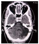
Background: Stroke is a common cause of morbidity and mortality worldwide. Therapeutic decision. regarding its management requires accurate diagnosis of stroke type and exclusion of mimics. Computed tomography (CT) scan has been found to be the gold standard in distinguishing haemorrhagic from ischemic stroke.
AIM: The aim of this study is to evaluate the pattern of CT finding in stroke patients in University of Port Harcourt Teaching Hospital and assess the accuracy of clinical assessment of stroke pattern.
Method: A retrospective review of computed tomography findings in 68 consecutive patients who were referred from different private clinics and medical ward of UPTH to the Radiology department with clinical diagnosis of stroke was undertaken over a period of one year (April 2007-March 2008). Age, sex, clinical diagnosis, CT finding and the interval between ictus and CT scan were recorded. All the CT examinations were performed on GE Hispeed-NX/1 Base-2002 Dual slice helical CT. Evaluation was done by consultant radiologists in the department.
Results: A total of 68 patients were scanned for stroke evaluation during the period under review, 37(54.4%) males and 31(45.6%) females, giving a male: female ratio of 1.6:1. The age range was 29-84. The mean interval between ictus and CT scan was 9 days. Nine patients had normal CT findings. Stroke patients were 48 (70.6%), 38(79.2%) were cerebral infarction while 10(20.8%) were intracerebral
heamorrhage. 2 (2.9%) patients had generalized cerebral edema, 1(2.2%) patient had brain tumour and 8 (11.8%) had generalized cerebral atrophy.
Conclusion: Evaluation of stroke pattern in this study shows that infarction is the commoner form of stroke subtype in this study. Stroke mimics were few. Stroke patients presented late and clinical differentiation of stroke subtypes are not reliable. Key words: computed tomography, hemorrhage, infarction, stroke
Downloads
Article Details
Section

This work is licensed under a Creative Commons Attribution-NonCommercial 4.0 International License.
This is an open access journal, and articles are distributed under the terms of the Creative Commons Attribution-NonCommercial-ShareAlike 4.0 License, which allows others to remix, tweak, and build upon the work non-commercially, as long as appropriate credit is given and the new creations are licensed under the identical terms.
How to Cite
References
1. Murray CJ, Lopez AD. Mortality by cause for eight regions of the world: global burden of disease study. Lancet 1997, 349;1269-1276
2. Stephen PK, Darius GN, Christian G et al. Acute Stroke Assemment with CT: Do we Need Multimodal Evaluation? Radiology 2004:23379-56
3. Mustapha D, Njideka O, Frank O Prevalence of stroke in an urban, mixed income community in Lagos, Nigeria. Neuroepidemiology 2007, 28-216-223
4. Ogungbo BL, Mendelow AD, Wakker R. Cerebrovascular diseases in Nigeria: what do we know and what do we need to know? Tropical Doctor 2003, 33:25-30
5. Osuntokun BO Stroke in the Africans. Atr. 1 Med. Sci. 1977,6:39-53
6. Ogun SA, Oluwole O, Ogunsevinde AG et al. Misdiagnosis of stroke-A computerized Tomography scan study. WAJM 2000, 19:19-22
7. Harold PA, Robert JA, Thomas B et al Guidelines for the early management of patients with ischemic stroke. A Scientific 30 statement from the stroke council of the American stroke association. Stroke 2003; 34: 1056-1053.
8. Weir CI, Murray GD), Adams FG et al. Poor accuracy of stroke scoring systems for differential clinical diagnosis of intracranial hemorrhage and infarction. Lancet 1994;344 999-1002.
9. Oreren A, Bicakci 5, Burgut R., Sarica Y Bozdemir H. Accuracy of bedside diagnosis versus Allen and Siriraj stroke scores in Turkish patients. European Journal of Neurology 2006;13:611-615.
10 Arbin M. Britton M, Faire U Helmers C Miah K. Murray V Accuracy of bedside diagnosis in stroke. Stroke 1961, 12:288-293
11. Januel AC, Tailleur T. Loubes-Lacroix F, et al. Imaging of cerebral ischemia within first hours.computed tomography (CT).Radiol 2005; 86: 1091-1101
12. Köniq M. Brain perfusion CT in acute stroke current status. Eur J. Radiol 2003, 45:11-22
13. James DE, Micheal HL, Max winter M, et al. Correlation of early dynamic CT perfusion imaging with whole brain MK diffusion and perfusion imaging in acute hemispheric stroke
14. Harold A, Robert A, Gregory DZ and Larry BG. Guidelines for the early management of patients with ischemic stroke 2005 guidelines update. A scientific statement from the stroke council of the American heart association/ American stroke association Stroke, 2005, 36:916-921
15. Komolafe MA, Komolafe ED, Fatove Fetal Profile of stroke in Nigeria: A prospective clinical study. African Journal of Neurological Sciences 2007:26:5-13
16. Kehinde OK, Shamsideen AO, Bamidele O Validation study of the Seriraj stroke score in African Nigeriars and evaluation of the discriminant values of its parameters, a preliminary prospective CT scan study. Stroke 2006, 37-1997-2000,
17. Bamford 1, Sandercock P, Dennis M, Burn 1. Warlow CA prospective study of acute cerebrovascular disease in the commsinity the Oxfordshire community stroke project 1951-86. 2. Incidences, case fatality rates and overall outcome
18. Nyame PK, Jumah KB, Adjei 5. Computerised tomographic scan of the head in evaluation of stroke in Ghanaians. East Afr Med 11998, 75:637-639
19. Odusote K. Management of stroke. Nigerian Medical Practitioner 1996: 32-56-62
20. Njoku CH, Aduloju AB Stoke in Sokoto, Nigeria: A five year retrospective study
21. Timmis GC Cardiovascular reviews William and Wilkins, Baltmore/ London 1950:266-267
22. Adams HP Jr. Brott TG, Furlan Al, et al Guidelines for thrombolytic therapy for acute stroke: a supplement to the guidelines for management of patients with acute ischemic stroke- a statement for healthcare professional from a special writing group of the stroke council, American Heart Association Stroke 1996 27-1711-1718