

Profile of computed tomography scan findings of patients diagnosed with pancreatic neoplasm at Dr. George Mukhari Academic Hospital, Ga‑Rankuwa, Pretoria, South Africa
Article Sidebar
Views | PDF/EPUB Downloads:
236
/ 56
/ 30
Main Article Content
Abstract
Background: The aim of this study is to determine the profile of abdominal computed tomography (CT) scan findings of patients diagnosed with pancreatic adenocarcinoma and other pancreatic neoplasm that simulates pancreatic adenocarcinoma, which constitute the majority of pancreatic neoplasm at Dr. George Mukhari Academic Hospital, Ga-Rankuwa, Pretoria, South Africa.
Materials and Methods: A prospective study of abdominal CT scans of 67 patients, aged 12 years and older, with histologically confirmed pancreatic neoplasms, including their medical records and laboratory results, from November 1, 2013 to June 30, 2017, was conducted. CT scan images were acquired with 128 slices, Philips, and GE CT scanners. Statistical analysis was made using a Statistical Program for the Social Sciences software SPSS (version 22.0).
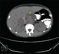
Results: There were 36 females(53.7%) and 31 males(46.3%) in this series and four demised. The ages of the patients ranged from 12 to 90 years. The most common clinical presentation was obstructive jaundice (86.6%). The predominant histological diagnosis was adenocarcinoma (74.6%), followed by primary lymphoma of the pancreas (13.4%) and 65.7% of the pancreatic neoplasms were unresectable, while most of the other pancreatic neoplasms based on their CT scan findings masqueraded as pancreatic adenocarcinoma. Pancreatic adenocarcinoma demonstrated both typical and atypical CT scan findings.
Conclusion: Accurate diagnosis and appropriate management of pancreatic neoplasms are important because of their high morbidity and mortality. The majority of the pancreatic neoplasms were unresectable at the time of their presentation. A multidisciplinary management team is recommended since pancreatic neoplasms still remain a serious clinical challenge.
Downloads
Article Details
Section

This work is licensed under a Creative Commons Attribution-NonCommercial 4.0 International License.
This is an open access journal, and articles are distributed under the terms of the Creative Commons Attribution-NonCommercial-ShareAlike 4.0 License, which allows others to remix, tweak, and build upon the work non-commercially, as long as appropriate credit is given and the new creations are licensed under the identical terms.
How to Cite
References
1. Anand MK, Karani J, Gupta N, Coombs BD, Schmiedl UP, Amin Z, et al. Pancreatic Adenocarcinoma Imaging. Available from:
http://www.emedicine.medscape.com/article/370909‑Overview. [Last accessed on 2015 Apr 02; Last retrieved on 2017 Jun 10].
2. Ryan DP, Hong TS, Bardeesy N. Pancreatic adenocarcinoma. N Engl J Med 2014;371:2140‑1.
3. Vincent A, Herman J, Schulick R, Hruban RH, Goggins M. Pancreatic cancer. Lancet 2011;378:607‑20.
4. Bond‑Smith G, Banga N, Hammond TM, Imber CJ. Pancreatic adenocarcinoma. BMJ 2012;344:e2476.
5. Alberts SR, Goldberg RM. Gastrointestinal cancer. In: Casciato DA, Territo MC, editors. Manual of Clinical Oncology. Ch. 9. Baltimore, USA: Lippincott Williams and Wilkins; 2009. p. 188‑236.
6. World Cancer Report. Ch. 5.7. World Health Organization. Available from: http://www.shop.iarc.fr/products/wcr2014. [Last retrieved
2017 Jun 10].
7. Bosetti C, Lucenteforte E, Silverman DT, Petersen G, Bracci PM, Ji BT, et al. Cigarette smoking and pancreatic cancer: An analysis from the
international pancreatic cancer case‑control consortium (Panc4). Ann Oncol 2012;23:1880‑8.
8. Wolfgang CL, Herman JM, Laheru DA, Klein AP, Erdek MA, Fishman EK, et al. Recent progress in pancreatic cancer. CA Cancer J Clin 2013;63:318‑48.
9. Yang MJ, Li S, Liu YG, Jiao N, Gong JS. Common and unusual CT and MRI manifestations of pancreatic adenocarcinoma: A pictorial review. Quant Imaging Med Surg 2013;3:113‑20.
10. Coakley FV, Hanley‑Knutson K, Mongan J, Barajas R, Bucknor M, Qayyum A, et al. Pancreatic imaging mimics: Part 1, imaging mimics of pancreatic adenocarcinoma. AJR Am J Roentgenol 2012;199:301‑8.
11. Ryan DP, Hong TS, Bardeesy N. Pancreatic adenocarcinoma. N Engl J Med 2014;371:1039‑49.
12. Mergo PJ, Helmberger TK, Buetow PC, Helmberger RC, Ros PR. Pancreatic neoplasms: MR imaging and pathologic correlation.
Radiographics 1997;17:281‑301.
13. Martin DR, Semelka RC. MR imaging of pancreatic masses. Magn Reson Imaging Clin N Am 2000;8:787‑812.
14. Ros PR, Mortelé KJ. Imaging features of pancreatic neoplasms. JBR‑BTR 2001;84:239‑49.
15. Low G, Panu A, Millo N, Leen E. Multimodality imaging of neoplastic and nonneoplastic solid lesions of the pancreas. Radiographics
2011;31:993‑1015.
16. Xu M, Sethi A. Imaging of the pancreas. Gastroenterol Clin 2016;45:101‑16.
17. Botcha S, Rangasami R, Johson T, Rajamanickam BS. Multidetector CT findings of pancreatic neoplasms with histopahatological correlation:
A pictorial essay. Int Surg J 2015;2:141‑6