

Preoperative dual imaging evaluation of profound sensorineural hearing loss in patients for cochlear implantation
Article Sidebar
Views | PDF/EPUB Downloads:
467
/ 99
/ 39
Main Article Content
Abstract
Background: Profound sensorineural hearing loss (SNHL) may be the result of major inner ear structural malformations, and cochlear implantation remains the only viable treatment option. High-resolution computed tomography (HRCT) and magnetic resonance imaging (MRI) are indispensable for optimum preoperative implant workup and thus play a vital role in patient selection, pre-implantation counseling, and surgical management.
Aim and Objectives: The aim of this study is to evaluate patients with profound SNHL for cochlear implantation preoperatively on both HRCT and MRI and to compare imaging findings in both modalities.
Materials and Methods: This longitudinal prospective study was conducted in the Department of Radiology of a tertiary care-based hospital in North India. A total of 45 patients (90 temporal bones) with clinically diagnosed bilateral profound SNHL were included in the study. Patients with a previous history of temporal bone injury were excluded from the study. All cases were evaluated on both 128 slice Philips computed tomography (CT) machine and 1.5 Tesla Siemens Magnetom MRI scanner. Each temporal bone was systematically analyzed for anatomical and structural abnormalities.
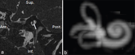
Results: Both high-resolution CT and MRI played vital roles in the workup of patients with profound SNHL for cochlear implantation and allowed accurate assessment of critical inner ear abnormalities. Cochlear malformations (30%) were responsible for the majority of structural abnormalities in this study with Type II incomplete partition (8.9%) being the most common. Cochlear nerve deficiency was seen in 20 cases (22.2%) and was diagnosed only on MRI. Similarly, early fibrosis and abnormal signal intensity were also detected
only on MRI, which were missed on CT.
Conclusions: Both high-resolution CT and high magnet MRI complement each other and reduce the chances of missing critical findings, which are crucial for surgical management and planning. Thus, it is advisable to perform dual imaging with both modalities wherever and whenever possible, to offer maximum information to treating surgeon preoperatively.
Downloads
Article Details
Section
This is an open access journal, and articles are distributed under the terms of the Creative Commons Attribution-NonCommercial-ShareAlike 4.0 License, which allows others to remix, tweak, and build upon the work non-commercially, as long as appropriate credit is given and the new creations are licensed under the identical terms.
How to Cite
References
1. Digge P, Solanki RN, Shah DC, Vishwakarma R, Kumar S. Imaging modality of choice for pre‑operative cochlear imaging: HRCT vs. MRI
temporal bone. J Clin Diagn Res 2016;10:TC01‑4.
2. Jackler RK, Luxford WM, House WF. Congenital malformations of the inner ear: A classification based on embryogenesis. Laryngoscope
1987;97:2‑14.
3. Young JY, Ryan ME, Young NM. Preoperative imaging of sensorineural hearing loss in pediatric candidates for cochlear implantation.
Radiographics 2014;34:E133‑49.
4. Joshi VM, Navlekar SK, Kishore GR, Reddy KJ, Kumar EC. CT and MR imaging of the inner ear and brain in children with congenital sensorineural hearing loss. Radiographics 2012;32:683‑98.
5. Taha T, Wahba H, Ibrahim AS, Abd Elazim Y. Cochlear implant tailored imaging protocol: What clinicians need to know. Egypt J Radiol Nucl
Med 2015;46:33‑43.
6. Bhavana K, Kumar S. Modifying radiology protocols for cochlear implant surgery in a government sponsored scheme: Need of the
hour. Int J Med Res Health Sci 2016;5:151‑7.
7. Demirpolat G, Savaş R, Totan S, Bilgen I, Kirazli T, Alper H. Temporal bone CT and MRI in cochlear implant candidates. Tani Girisim Radyol
2003;9:41‑6.
8. Aschendorff A. Imaging in cochlear implant patients. GMS Curr Top Otorhinolaryngol Head Neck Surg 2011;10:Doc07.
9. Huang BY, Zdanski C, Castillo M. Pediatric sensorineural hearing loss, part 1: Practical aspects for neuroradiologists. AJNR Am J Neuroradiol
2012;33:211‑7.
10. Gupta SS, Maheshwari SR, Kirtane MV, Shrivastav N. Pictorial review of MRI/CT Scan in congenital temporal bone anomalies, in patients
for cochlear implant. Indian J Radiol Imaging 2009;19:99‑106.
11. Connor SE, Bell DJ, O’Gorman R, Fitzgerald‑O’Connor A. CT and MR imaging cochlear distance measurements may predict cochlear implant length required for a 360 degrees insertion. AJNR Am J Neuroradiol 2009;30:1425‑30.
12. Roche JP, Huang BY, Castillo M, Bassim MK, Adunka OF, Buchman CA. Imaging characteristics of children with auditory neuropathy spectrum disorder. Otol Neurotol 2010;31:780‑8.
13. Sennaroglu L, Saatci I. A new classification for cochleovestibular malformations. Laryngoscope 2002;112:2230‑41.
14. Chaturvedi A, Mohan C, Mahajan S, Kakkar V. Imaging of cochlear implants. Indian J Radiol Imag 2006;16:385‑92.
15. Mazón M, Pont E, Montoya‑Filardi A, Carreres‑Polo J, Más‑Estellés F. Inner ear malformations: A practical diagnostic approach. Radiologia
2017;59:297‑305.
16. Verbist BM. Imaging of sensorineural hearing loss: A pattern‑based approach to diseases of the inner ear and cerebellopontine angle.
Insights Imaging 2012;3:139‑53.
17. Phelps PD. Cochlear implants for congenital deformities. J Laryngol Otol 1992;106:967‑70.
18. Verbist BM, Frijns JH, Geleijns J, van Buchem MA. Multisection CT as a valuable tool in the postoperative assessment of cochlear implant
patients. AJNR Am J Neuroradiol 2005;26:424‑9.
19. Gomes ND, Couto CL, Gaiotti JO, Maria A, Costa D, Ribeiro MA, et al. Cochlear implant: What the radiologist should know. Radiol Bras
2013;46:163‑7.
20. Witte RJ, Lane JI, Driscoll CL, Lundy LB, Bernstein MA, Kotsenas AL, et al. Pediatric and adult cochlear implantation. Radiographics 2003; 23: 1185‑200.
21. Alam‑Eldeen MH, Rashad UM, Ali AH. Radiological requirements for surgical planning in cochlear implant candidates. Indian J Radiol
Imaging 2017; 27:274‑81.
22. Cremers CW, van Rijn PM, Huygen PL. The sex‑ratio in childhood deafness, an analysis of the male predominance. Int J Pediatr
Otorhinolaryngol 1994;30:105‑10.
23. Bamiou DE, Phelps P, Sirimanna T. Temporal bone computed tomography findings in bilateral sensorineural hearing loss. Arch Dis Child 2000;82:257‑60.
24. Yiin RS, Tang PH, Tan TY. Review of congenital inner ear abnormalities on CT temporal bone. Br J Radiol 2011;84:859‑63.
25. Westerhof JP, Rademaker J, Weber BP, Becker H. Congenital malformations of the inner ear and the vestibulocochlear nerve in children with sensorineural hearing loss: Evaluation with CT and MRI. J Comput Assist Tomogr 2001;25:719‑26.
26. Adunka OF, Roush PA, Teagle HF, Brown CJ, Zdanski CJ, Jewells V, et al. Internal auditory canal morphology in children with cochlear nerve deficiency. Otol Neurotol 2006;27:793‑801.
27. Komatsubara S, Haruta A, Nagano Y, Kodama T. Evaluation of cochlear nerve imaging in severe congenital sensorineural hearing loss. ORL J Otorhinolaryngol Relat Spec 2007;69:198‑202.
28. Ellul S, Shelton C, Davidson HC, Harnsberger HR. Preoperative cochlear implant imaging: Is magnetic resonance imaging enough?
Am J Otol 2000;21:528‑33.
29. Bath AP, O’Donoghue GM, Holland IM, Gibbin KP. Paediatric cochlear implantation: How reliable is computed tomography in assessing cochlear patency? Clin Otolaryngol Allied Sci 1993;18:475‑9.
30. Seitz J, Held P, Waldeck A, Strotzer M, Völk M, Strutz J, et al. Value ofhigh‑resolution MR in patients scheduled for cochlear implantation.
Acta Radiol 2001;42:568‑73.