

Intracranial Cystic Tumors with a Mural Nodule: Conventional, Diffusion Tensor and Perfusion Magnetic Resonance Imaging Findings
Article Sidebar
Views | PDF/EPUB Downloads:
71
/ 9
Main Article Content
Abstract
Introduction: Intracranial cystic tumors with mural nodule have been extensively studied. None of these studies have established the
role of conventional, fractional anisotropy diffusion tensor imaging (FADTI) and dynamic susceptibility contrast magnetic resonance
imaging (MRI) in the assessment of grade and type of cystic brain tumors with a mural nodule.
Materials and Methods: This was a retrospective cross-sectional study, which was conducted at Severance Hospital. Brain MRI of 15 consecutive patients with a cystic brain tumor with a mural nodule was analyzed.
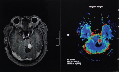
Results: Among the 15 studied patients 11 were females. The age ranged from 12 to 54 years. Six of the patients had high‑grade tumors. Most five of the high‑grade tumors showed cystic wall contrast enhancement while none was found among the low-grade group. All high-grade tumors showed increased regional cerebral blood volume (rCBV) compared to three of low-grade tumors. Hemangioblastomas, glioblastoma multiforme (GBM) and the primitive neuroectodermal tumor showed increased rCBV while pilocytic astrocytoma, ependymoma, and ganglioglioma showed decreased rCBV. There were no difference in fractional anisotropy (FA) values between tumor grades and types.
Conclusion: Postcontrast T1-weighted image and perfusion MRI showed in this study to be very useful in differentiating high- and low-grade cystic tumors with the mural nodule. FA values added no benefit to tumor differentiation. Hemangioblastoma was the only tumor with increased rCBV among low‑grade tumors. GBM, which is a malignant tumor, can present as a cystic lesion with a mural nodule.
Downloads
Article Details
Section

This work is licensed under a Creative Commons Attribution-NonCommercial 4.0 International License.
This is an open access journal, and articles are distributed under the terms of the Creative Commons Attribution-NonCommercial-ShareAlike 4.0 License, which allows others to remix, tweak, and build upon the work non-commercially, as long as appropriate credit is given and the new creations are licensed under the identical terms.
How to Cite
References
1. Rollin N, Guyotat J, Streichenberger N, Honnorat J, Tran Minh VA, Cotton F. Clinical relevance of diffusion and perfusion magnetic resonance imaging in assessing intra‑axial brain tumors. Neuroradiology 2006;48:150‑9.
2. Pierpaoli C, Basser PJ. Toward a quantitative assessment of diffusion anisotropy. Magn Reson Med 1996;36:893‑906.
3. Bammer R, Augustin M, Strasser‑Fuchs S, Seifert T, Kapeller P, Stollberger R, et al. Magnetic resonance diffusion tensor imaging for characterizing diffuse and focal white matter abnormalities in multiple sclerosis. Magn Reson Med 2000;44:583‑91.
4. Ciccarelli O, Werring DJ, Wheeler‑Kingshott CA, Barker GJ, Parker GJ, Thompson AJ, et al. Investigation of MS normal‑appearing brain using diffusion tensor MRI with clinical correlations. Neurology 2001;56:926‑33.
5. Ellis CM, Simmons A, Jones DK, Bland J, Dawson JM, Horsfield MA, et al. Diffusion tensor MRI assesses corticospinal tract damage in ALS. Neurology 1999;53:1051‑8.
6. Hakyemez B, Erdogan C, Ercan I, Ergin N, Uysal S, Atahan S. High‑grade and low‑grade gliomas: Differentiation by using perfusion MR imaging. Clin Radiol 2005;60:493‑502.
7. Sugahara T, Korogi Y, Shigematsu Y, Liang L, Yoshizumi K, Kitajima M, et al. Value of dynamic susceptibility contrast magnetic resonance imaging in the evaluation of intracranial tumors. Top Magn Reson Imaging 1999;10:114‑24.
8. Shin JH, Lee HK, Kwun BD, Kim JS, Kang W, Choi CG, et al. Using relative cerebral blood flow and volume to evaluate the histopathologic grade of cerebral gliomas: Preliminary results. AJR Am J Roentgenol 2002;179:783‑9.
9. Di Costanzo A, Pollice S, Trojsi F, Giannatempo GM, Popolizio T, Canalis L, et al. Role of perfusion‑weighted imaging at 3 Tesla in the assessment of malignancy of cerebral gliomas. Radiol Med 2008;113:134‑43.
10. Law M, Kazmi K, Wetzel S, Wang E, Iacob C, Zagzag D, et al. Dynamic susceptibility contrast‑enhanced perfusion and conventional MR imaging findings for adult patients with cerebral primitive neuroectodermal tumors. AJNR Am J Neuroradiol 2004;25:997‑1005.
11. Bing F, Kremer S, Lamalle L, Chabardes S, Ashraf A, Pasquier B, et al. Value of perfusion MRI in the study of pilocytic astrocytoma and hemangioblastoma: Preliminary findings. J Neuroradiol 2009;36:82‑7.
12. Maiuri F. Cysts with mural tumor nodules in the cerebral hemispheres. Neurosurgery 1988;22:703‑6.
13. Maiuri F, Stella L, Benvenuti D, Gangemi M, Corriero G. Tumors of the neuron series presenting as cysts with mural nodules. Acta Neurol (Napoli) 1989;11:25‑31.
14. Sridhar K, Ravi R, Ramamurthi B, Vasudevan MC. Cystic meningiomas. Surg Neurol 1995;43:235‑9.
15. Dwarakanath S, Suri A, Sharma BS, Mehta VS. Intracranial hemangioblastomas: An institutional experience. Neurol India 2006;54:276‑8.
16. Weerakkody FG. Pilocytic Astrocytoma | Radiology Reference Article | Radiopaedia.org. Available from: http://www.radiopaedia.org/articles/pilocytic‑astrocytoma. [Last cited on 2014 Nov 17].
17. Osborn AG. Astrocytomas and other glial neoplasms. Diagnostic Neuroradiology. Mosby: St. Louis, 1994. p. 529‑78.
18. Fernandez C, Figarella‑Branger D, Girard N, Bouvier‑Labit C, Gouvernet J, Paz Paredes A, et al. Pilocytic astrocytomas in children: Prognostic factors – A retrospective study of 80 cases. Neurosurgery 2003;53:544‑53.
19. Sinha S, Bastin ME, Whittle IR, Wardlaw JM. Diffusion tensor MR imaging of high‑grade cerebral gliomas. AJNR Am J Neuroradiol 2002;23:520‑7.
20. Sakamoto T, Sakakibara Y, Hayashi T, Yamashita K, Sekino H, Ozawa T, et al. Recurrence of pleomorphic xanthoastrocytoma six years after total removal of mural nodule: A case report. No Shinkei Geka 1995;23:941‑5.
21. Maldaun MV, Suki D, Lang FF, Prabhu S, Shi W, Fuller GN, et al. Cystic glioblastoma multiforme: Survival outcomes in 22 cases. J Neurosurg 2004;100:61‑7.
22. Tsuchiya K, Fujikawa A, Nakajima M, Honya K. Differentiation between solitary brain metastasis and high‑grade glioma by diffusion tensor imaging. Br J Radiol 2005;78:533‑7.
23. Osborn AG, Salzman KL, Barkovich J. Neoplasms, Diagnostic Imaging Brain. 2nd ed. Friesens: Altona, 2010. p. I: 6‑82.
24. Calli C, Kitis O, Yunten N, Yurtseven T, Islekel S, Akalin T. Perfusion and diffusion MR imaging in enhancing malignant cerebral tumors. Eur J Radiol 2006;58:394‑403.
25. Métellus P, Ait Ameur A, Faillot T, Camparo P, Dutertre G, Desgeorges M. Contribution of perfusion magnetic resonance imaging in a patient with cerebral ganglioglioma. Neurochirurgie 2002;48:426‑30.
26. Filippi M, Cercignani M, Inglese M, Horsfield MA, Comi G. Diffusion tensor magnetic resonance imaging in multiple sclerosis. Neurology 2001;56:304‑11.
27. Tievsky AL, Ptak T, Farkas J. Investigation of apparent diffusion coefficient and diffusion tensor anisotrophy in acute and chronic multiple sclerosis lesions. AJNR Am J Neuroradiol 1999;20:1491‑9.
28. Hajnal JV, Doran M, Hall AS, Collins AG, Oatridge A, Pennock JM, et al. MR imaging of anisotropically restricted diffusion of water in the nervous system: Technical, anatomic, and pathologic considerations. J Comput Assist Tomogr 1991;15:1‑18.
29. Wieshmann UC, Clark CA, Symms MR, Franconi F, Barker GJ, Shorvon SD. Reduced anisotropy of water diffusion in structural cerebral abnormalities demonstrated with diffusion tensor imaging. Magn Reson Imaging 1999;17:1269‑74.
30. Inoue T, Ogasawara K, Beppu T, Ogawa A, Kabasawa H. Diffusion tensor imaging for preoperative evaluation of tumor grade in gliomas. Clin Neurol Neurosurg 2005;107:174‑80.
31. Tropine A, Vucurevic G, Delani P, Boor S, Hopf N, Bohl J, et al. Contribution of diffusion tensor imaging to delineation of gliomas and glioblastomas. J Magn Reson Imaging 2004;20:905‑12.
32. Lee HY, Na DG, Song IC, Lee DH, Seo HS, Kim JH, et al. Diffusion‑tensor imaging for glioma grading at 3‑T magnetic resonance imaging: Analysis of fractional anisotropy and mean diffusivity. J Comput Assist Tomogr 2008;32:298‑303.
33. Goebell E, Paustenbach S, Vaeterlein O, Ding XQ, Heese O, Fiehler J, et al. Low‑grade and anaplastic gliomas: Differences in architecture evaluated with diffusion‑tensor MR imaging. Radiology 2006;239:217‑22.
34. White ML, Zhang Y, Yu F, Jaffar Kazmi SA. Diffusion tensor MR imaging of cerebral gliomas: Evaluating fractional anisotropy characteristics. AJNR Am J Neuroradiol 2011;32:374‑81.
35. Liu X, Tian W, Kolar B, Yeaney GA, Qiu X, Johnson MD, et al. MR diffusion tensor and perfusion‑weighted imaging in preoperative grading of supratentorial nonenhancing gliomas. Neuro Oncol 2011;13:447‑55.