

An observational study of the demographic, clinical, and diffusion‑weighted magnetic resonance imaging characteristics of patients with musculoskeletal infections
Article Sidebar
Views | PDF/EPUB Downloads:
439
/ 97
/ 40
Main Article Content
Abstract
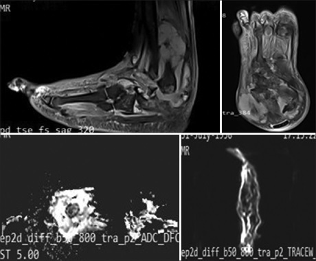
Introduction: Musculoskeletal infections have been emerging nowadays. Its early diagnosis is warranted as it may lead to disabling sequelae. Recently, the use of diffusion-weighted magnetic resonance imaging (DWMRI) provided additional pulse sequences enabling better diagnosis and needs to be explored for diagnosing musculoskeletal infections. Thus, we conducted this study with an aim to discuss demographic, clinical, and DWMRI findings of the spectrum of musculoskeletal infections, emphasizing the apparent diffusion
coefficient (ADC) map for this domain of infections.
Methods: A retrospective observational study was carried out in the department of radiodiagnosis of a tertiary care hospital. The study was performed on 50 patients who were suspected cases of musculoskeletal infections. All the patients underwent basic investigations, ultrasound, magnetic resonance imaging, and diffusion-weighted imaging with ADC mapping. The data were entered into MS EXCEL spreadsheet and analysis was done using Statistical Package for Social Sciences (SPSS) version 21.0.
Results: Maximum patients were in the age group of 11–20 years (40%) with 58% males and 42% females. Lower limb infections were common, especially the involvement of the hip joint. Pain and swelling were the most common symptoms as seen in 96% and 88% of the patients respectively. DWMRI was able to diagnose and lay down significantly different ADC values for different musculoskeletal infections. The mean ADC values were higher for acute infections and lower for chronic infections.
Conclusions: DWMRI holds an important role in the investigation profile for musculoskeletal infections and must be used wherever deemed necessary to avoid unnecessary referrals and treatments.
Downloads
Article Details
Section
This is an open access journal, and articles are distributed under the terms of the Creative Commons Attribution-NonCommercial-ShareAlike 4.0 License, which allows others to remix, tweak, and build upon the work non-commercially, as long as appropriate credit is given and the new creations are licensed under the identical terms.
How to Cite
References
1. Stalcup ST, Hughes TH, Pathria MN. Musculoskeletal infections of the extremities: A tour from superficial to deep. Appl Radiol 2011;40:12‑22.
2. Dean Deyle G. The role of MRI in musculoskeletal practice: A clinical perspective. J Man Manip Ther 2011;19:152‑61.
3. Soldatos T, Durand DJ, Subhawong TK, Carrino JA, Chhabra A. Magnetic resonance imaging of musculoskeletal infections: Systematic diagnostic assessment and key points. Acad Radiol 2012;19:1434‑43.
4. Lalam RK, Cassar‑Pullicino VN, Tins BJ. Magnetic resonance imaging of appendicular musculoskeletal infection. Top Magn Reson Imaging 2007;18:177‑91.
5. Kumar Y, Khaleel M, Boothe E, Awdeh H, Wadhwa V, Chhabra A. Role of diffusion weighted imaging in musculoskeletal infections: Current perspectives. Eur Radiol 2017;27:414‑23.
6. Okubo Y, Nochioka K, Testa M. Nationwide survey of pediatric acute osteomyelitis in the USA. J Pediatr Orthop B 2017;26:501‑6.
7. Romeih M, Raafat T, Khalaf M, Sallam K. The diagnostic value of diffusion‑weighted magnetic resonance imaging in characterization
of musculoskeletal soft tissue tumors. Egypt J Radiol Nucl Med 2018;49:400‑7.
8. Kim J, Lee MU, Kim TH. Nationwide epidemiologic study for pediatric osteomyelitis and septic arthritis in South Korea: A cross‑sectional study of national health insurance review and assessment service. Medicine (Baltimore) 2019;98:e15355.
9. Jaramillo D, Dormans JP, Delgado J, Laor T, St. Geme JW 3 rd. Hematogenous osteomyelitis in infants and children: Imaging of a changing disease. Radiology 2017;283:629‑43.
10. Golsha R, Mehravar F, Alinezhad Esboie A, Rafiee S, Rafiee S. The epidemiology of skeletal tuberculosis in Northeast of Iran: A review of 229 cases. Iran J Med Sci 2018;43:380‑5.
11. Pati S, De S, Ghosh TN, Ghosh MK. Multifocal pure tubercular osteomyelitis: An unusual presentation in childhood. Indian J Tuberc 2017; 64:136‑40.