

Right Renal Pelvic Calculus Mimicking an Extrarenal Pelvis
Article Sidebar
Views | PDF/EPUB Downloads:
230
/ 31
/ 23
Main Article Content
Abstract
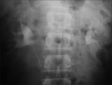
A 57-year-old man presented with recurrent intermittent colicky right flank pain of 1-year duration. Intravenous urogram (IVU) on two separate occasions suggested a right-sided, extrarenal pelvis. However, when pain became recurrent and persistent, he had a non-contrast computed tomography (CT) examination, which revealed a calculus in the renal pelvis. Diagnosis was missed in the initial imaging modalities because apart from the dilated pelvis, there was no evidence of hydronephrosis or calculus seen, hence a diagnosis of extrarenal pelvis. This case report highlights the superior utility of CT in imaging of suspected urolithiasis, especially when the patient remains symptomatic. Radiologists should be wary, especially in symptomatic patients with features of extrarenal pelvis on IVU.
Downloads
Article Details
Section

This work is licensed under a Creative Commons Attribution-NonCommercial 4.0 International License.
This is an open access journal, and articles are distributed under the terms of the Creative Commons Attribution-NonCommercial-ShareAlike 4.0 License, which allows others to remix, tweak, and build upon the work non-commercially, as long as appropriate credit is given and the new creations are licensed under the identical terms.
How to Cite
References
1. Sheafor DH, Hertzberg BS, Freed KS, Carroll BA, Keogan MT, Paulson EK et al. Nonenhanced helical CT and US in the emergency
evaluation of patients with renal colic: Prospective comparison. Radiology 2000;217:792‑7.
2. Tamm EP, Silverman PM, Shuman WP. Evaluation of the patient with flank pain and possible ureteral calculus. Radiology 2003; 228:319‑29.
3. Fowler KA, Locken JA, Duchesne JH, Williamson MR. US for detecting renal calculi with nonenhanced CT as a reference standard. Radiology 2002;222:109‑13.
4. Chen MY, Zagoria RJ. Can noncontrast helical computed tomography replace intravenous urography for evaluation of patients with acute urinary tract colic? J Emerg Med 1999;17:299‑303.
5. Dunnick NR, Sandler CM, Newhouse JH. Textbook of uroradiology. 3rd ed. Philadelphia: Lippincott Williams and Wilkins; 2001.
p. 22‑66.
6. Cheng PM, Moin P, Dunn MD, Boswell WD, Duddalwar VA. What the Radiologist Needs to Know About Urolithiasis: Part 2‑CT
Findings, Reporting, and Treatment. AJR Am J Roentgenol 2012;198:W548‑54.
7. Shih WJ, Lorman JJ, King JJ. Extrarenal pelvis mimicking obstructive uropathy in nine years of serial bone scintigraphies. Radiat Med 1998;6:253‑5.
8. Smith RC, Rosenfield AT, Choe KA, Essenmacher KR, Verga M, Glickman MG, et al. Acute flank pain: Comparison of non‑contrast‑enhanced CT and intravenous urography. Radiology 1995;194:789‑94.
9. Fielding JR, Steele G, Fox LA, Heller H, Loughlin KR. Spiral computerized tomography in the evaluation of acute flank pain: A replacement for excretory urography. J Urol 1997;157:2071‑3.
10. Levine JA, Neitlich J, Verga M, Dalrymple N, Smith RC. Ureteral calculi in patients with flank pain: Correlation of plain radiography
with unenhanced helical CT. Radiology 1997;204:27‑31.
11. Furlan A, Federle MP, Yealy DM, Averch TD, Pealer K. Nonobstructing renal stones on unenhanced CT: A Real Cause for Renal Colic? AJR Am J Roentgenol 2008;190:W125‑7.