

Ultrasound Evaluation Of Normal Liver Size And Factors Affecting It Among Adults In Northeastern Nigeria
Article Sidebar
Views | PDF/EPUB Downloads:
123
/ 59
Main Article Content
Abstract
Background: A large number of pat buloge satitica curs affect the sizes of the ver, and fimical cosmination is far fа мехри cletect small increases wie A previne study had observed that the liver size awer in Zimbabweans cumpared to Germana due to стесні Іспpiral diseases Thes stuttly was carried out in adults to find out the smaller se would alse he Jocuriented among a Nigerian population.
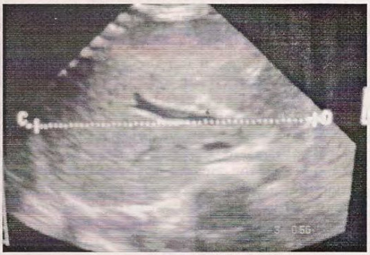
Methodology: The eite of the liver was mperated using ultrasoune scanning minkine fitted with 3.5MHe transducer in aduite aged between 18 arul fish yvire This prospeutive study was conducted in 106 Temales and U2 males with normal inver sonogrαρδώς πυδdinge The gittal und transverse diameters were measured and tichuted to ape, sex weight and height of the
Results: The averall mean and standard deviatino (80) of the liver size in sartal and ples were 0.35 cm Ling saf 13.73 cm + 0.19) zespectively. The sagitial liver span m the mod-clantcular lime was smaller sonng Nigerians when compared with the previous studies song Caucastana and Turkish indigeue A significent statistical difference ins0 05 wan abserved in the uunsenise liver span be wont male and female subjects, howmer, no significant stistical difference was abserved in the sagittal dimension. The nen, body weighit and height showed a weak positive correlation with the sound transverse inver dimerisices. Among motors, age showed the strongect correlation with the sagittal liver dirunssian and wolcht with the transverse liver dimen
Conclusion: The sagitral liver span in the mid-clavicular line from this study was amalier than valuse documented from the previous stucies on Caucasians and Turkish andigenes Subject age showed the strongest positive nervulacion with the sagittal liver sizes and weight with the transverse liver size
Downloads
Article Details
Section

This work is licensed under a Creative Commons Attribution-NonCommercial 4.0 International License.
This is an open access journal, and articles are distributed under the terms of the Creative Commons Attribution-NonCommercial-ShareAlike 4.0 License, which allows others to remix, tweak, and build upon the work non-commercially, as long as appropriate credit is given and the new creations are licensed under the identical terms.
How to Cite
References
1. Nafeh MA, Medhat A, Swifae Y, Moftah FM, Mohamed A, Soliman AG, et al. Ultrasounographic changes of the liver in Schistosoma haematobium infection. Am J Trop Med Hyg. 1992; 47 (2): 225-230.
2. Friis H, Ndhlovu P,Mdulasa T, Kaondera K, Franke D, Vennervald BJ, et al. Ultrasound organomegaly: liver and spleen dimensions among children in Zimbabwe. Trop Med Int Health. 1996; 1(2):183-190.
3. Ademoh DI. Prevalence of Shistosoma haematobium infection in parts of Yobe State of Nigeria. J Med Lab Sc. 1998; 7: 11-16.
4. Zoli M, Magalotti D, Grimaldi M, Gueli C, Marchesini G, Pisi E. Physical examination of the liver: is it still worth it? Am J Gastroenterol. 1995; 90 (9): 1428-32.
5. Kornus OL, Ozdemir A, Akkoya A, Erbas G, Celik H, Isike S. Normal liver, spleen and kidney dimensions- evaluation with sonography.AJR. 1998; 171:19.
6. Leoppold G, Hykes D. Imaging of the liver. In: Leopold G, Hykes D (eds). Ultrasound diagnosis. 3'd Edition. Churchill Livingstone. London. 1985; Pp 1-25.
7. Bartoon TJ, Brown BP, Abu-YousefMM, Ferguson KJ, Swhweiger GD, Erkonen WE, et al. Teaching physical RadiOl.1998;5(2): 101-3.
8. Brooks M. The Liver. In: Goldberg B, Petterson H (eds). Ultrasonography. The Nicer Institude. Oslo. 1996; Pp. 55-57.
9. Hagen WA. Real time ultrasound imaging in internal medicine. In: Hagen WA (eds). Ultasound atlas. l " Edition, Weintieim, London. 1985;
Pp25-221.
10. Bisset RAL, Khan AN. Liver, Billiary system, Spleen and Pancreas. In: Diffemtial Diagnosis in abdominal ultrasound Bisset RAL, Khan AN(eds). i" ed. Bailliare Tindal. London. 1991: Pp 22-23.
11. Kratzer W, Fritz V, Mason RA, Haenle MM, Kaechele V, Roemerstein S. Factors affecting liver size : a Sonographic survey of 2080 subjects.
J Ultrasound Med. 2003; 22(11): 1155-61.
12. Yazdanpanah Y,Thomas AK,Kardorff R, Talla I, Sow S, Niang M, et al. Organometric investigations of the spleen and liver by ultrasound in Schistosoma mansoni endemic and nonendemic villages in Senegal. Am J Trop Med Hyg. 1997; 57(2): 245-249.
13. Friis H, Ndhlovu P, Kaondera K, Franke D, Vennervald BJ, Christensen NO, et al. Ultrasounographic assessment of Schistosoma mansoni. and S haematobium morbidity in Zimbabwean Schoolchildren. Am J Trop Med Hyg. 1996; 55(3): 290-294.
14. Eltoum lA, Saad AM, Ismail BM, Ali MM, Suliaman S, Bennett JL, Homeida MA. Liver sonography in an area endemic for Schistosomiasis haematobium. Am J Trop Med Hyg. 1993; 48(1): 77-81.
15. Niederau C, Sonnenberg A, Muller JE, Erckenbrecht JF, Scholten T, Fritsch WP. Sonographic measurement of the normal liver, spleen, pancreas and portal vein. Radiology. 1983; 149(2): 537-540.