

Audit of pediatric computed tomography at Aminu Kano teaching hospital, Kano, nigeria
Article Sidebar
Views | PDF/EPUB Downloads:
222
/ 17
/ 23
Main Article Content
Abstract
Context: Computed tomography (CT) is becoming popular with advances in imaging technology, and pediatric imaging is also affected by this trend.
Aims: This study aimed to determine the pattern of pediatric CT scanning practice and common findings at Aminu Kano teaching hospital (AKTH).
Settings and Design: The study was conducted at the Radiology department of AKTH. It was a retrospective
descriptive study.
Materials and Methods: Patients aged between 4 days to 14 years and examined with a 4-slice Bright speed CT scanner at the Radiology department from January to December, 2011 were reviewed in this study. Information concerning the age,
gender, indications for the CT scan, type of CT scan conducted, and findings were recorded.
Statistical Analysis: Summarising indices were used (including frequencies, means, modes, and standard deviations).
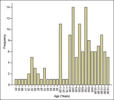
Results: One hundred and forty children (80 boys and 60 girls) were reviewed. Their ages ranged from 4.0 days to 14.0 years, with a mean of 5.64±4.31 years. Brain scan was most commonly performed (88.8%), while frequency of abdominal CT was 4.9%. The most common indication for CT examination in these subjects was convulsion (21.43%), followed by trauma (15.71%) and progressive head enlargement (11.43%). About 29.2% of the scans were normal, while obstructive hydrocephalus was seen in 13.2% and general brain atrophy in 9.1% of the cases.
Conclusions: This review shows predominance of brain CT scan in children, seizures and trauma being the most common indications. Obstructive hydrocephalus, brain infarction, and general atrophy dominate the findings. Presence of global atrophy in some of the patients is worrisome as it may adversely affect the prognosis.
Downloads
Article Details
Section

This work is licensed under a Creative Commons Attribution-NonCommercial 4.0 International License.
This is an open access journal, and articles are distributed under the terms of the Creative Commons Attribution-NonCommercial-ShareAlike 4.0 License, which allows others to remix, tweak, and build upon the work non-commercially, as long as appropriate credit is given and the new creations are licensed under the identical terms.
How to Cite
References
1. Brenner DJ, Hall EJ. Computed tomography — an increasing source of radiation exposure. N Engl J Med 2007;357:2277‑84.
2. Donnelly LF. Reducing radiation dose associated with pediatric CT by decreasing unnecessary examinations. AJR Am J Roentgenol
2005;184:655‑7.
3. United Nations Scientific Committee on Effects of Atomic Radiation. Sources and effects of ionizing radiation. UNSCEAR 2000, Report to the general assembly. vol. 1. New York; 2000.
4. Nzeh D, Oyinloye OI, Odebode OT, Akande H, Braimoh K. Ultrasound evaluation of brain infections and its complications in Nigerian infants. Trop Doct 2010;40:178‑80.
5. Jennet B. Epidemiology of head injury. Arch Dis Child 1998;78:403‑6.
6. Islam MN, Rasul CN, Sarder AH, Hossain SA. Computed tomographic evaluation of paediatric brain in a teaching hospital. Bang Med J (Khulna) 2011;44:3‑6.
7. Graham DI. Paediatric head injury. Brain 2001;124:1261‑2.
8. Suwaid MA, Tabari MA, Isyaku K, Idris SK, Saleh MK, Abdulkadir AY. Sonographic measurement of normal thyroid gland volume in school children in Kano Nigeria. West Afr J Ultrasound 2007;8:14‑22.
9. Idro R, Gwer S, Kahindi M, Gatakaa H, Kazungu T, Ndiritu M, et al. The incidence, aetiology and outcome of acute seizures in children admitted to a rural Kenyan district hospital. BMC Pediatr 2008;8:5.
10. Fenton SJ, Hansen KW, Meyers RL, Vargo DJ, White KS, Firth SD, et al. CT scan and the pediatric trauma patient—are we overdoing
it? J Pediatr Surg 2004;39:1877‑81.