

Normal ultrasonographic dimensions of the gallbladder and common bile duct in neonates
Article Sidebar
Views | PDF/EPUB Downloads:
513
/ 167
/ 40
Main Article Content
Abstract
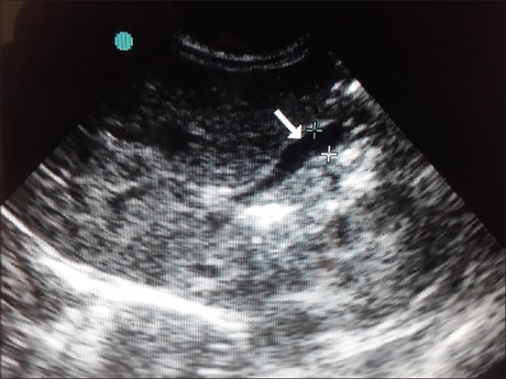
Background: Ultrasound (US) is the first choice of imaging in neonates presenting with persistent jaundice to exclude surgically correctable causes and differentiate obstructive from nonobstructive causes. Previous studies on normal dimensions of gallbladder (GB) and common bile duct (CBD) recruited adults and children spread across a wide age group.
Aims: This study aimed to determine GB and CBD normal dimensions in a large homogeneous neonatal population as well as guide decision regarding pre-US fasting in neonates who require GB evaluation.
Materials and Methods: Five hundred and twenty-eight healthy newborns were recruited between May 2009 and May 2011. The widest intraluminal anterior-posterior diameters of GB and CBD were measured. Neonatal age in days, sex, birth weight, weight and height, gestational age at delivery, and time interval since last feed recorded.
Results: The mean age was 9.56 ± 7.66 days, and 50.6% were males. The mean CBD diameter was 1.16 ± 1.61 mm while the mean GB diameter was 4.42 ± 2.16 mm. GB and CBD were clearly seen and measurable in 297 (55.8%) neonates and 237 (44.38%) neonates, respectively. There was a significant correlation between CBD diameter and GB diameter (P = 0.04) but no correlation with any demographic parameter. GB visualization was not dependent on time interval from last feed.
Conclusion: Mean neonatal values for CBD and GB were established, but neonates have a wider range of GB diameters compared with older children, so GB diameter may not be a reliable parameter for neonatal GB pathologies. GB visualization was not dependent on time interval from last feed; hence, a recent feed should not delay emergency scans, especially in ill neonates
Downloads
Article Details
Section
This is an open access journal, and articles are distributed under the terms of the Creative Commons Attribution-NonCommercial-ShareAlike 4.0 License, which allows others to remix, tweak, and build upon the work non-commercially, as long as appropriate credit is given and the new creations are licensed under the identical terms.
How to Cite
References
1. Hernanz‑Schulman M, Ambrosino MM, Freeman PC, Quinn CB. Common bile duct in children: Sonographic dimensions. Radiology 1995; 195:193‑5.
2. Glazer G, Filly R, Laing F. Rapid change in calibre of the non‑obstructed common duct. Radiology 1981;140:161‑2.
3. Simeone JF, Butch RJ, Mueller PR, vanSonnenberg E, Ferrucci JT Jr, Hall DA, et al. The bile ducts after a fatty meal: Further sonographic
observations. Radiology 1985;154:763‑8.
4. Cooperberg PL, Li D, Wong P, Cohen MM, Burhenne HJ. Accuracy of common hapatic duct size in the evaluation of extrahepatic biliary obstruction. Radiology 1980;135:141‑4.
5. Jaw T S, Kuo YT, Liu GC, Chen SH, Wang CK. MR cholangiography in the evaluation of neonatal cholestasis. Radiology 1999; 212:249‑56.
6. Sarin YK, Sengar M, Puri AS. Forme fruste choledochal cyst. Indian Pediatr 2005;42:1153‑5.
7. Wu CC, Ho YH, Chen CY. Effect of aging on common bile ductdiameter: A real‑time ultrasonographic study. J Clin Ultrasound 1984;12:473‑8.
8. Horrow MM, Horrow JC, Niakosari A, Kirby CL, Rosenberg HK. Is age associated with size of adult extrahepatic bile duct: Sonographic study. Radiology 2001;221:411‑4.
9. Lindholm EB, Meckmongkol T, Feinberg AJ, Kim A, Ciullo S, Mallon M, et al. Standardization of common bile duct size using ultrasound in pediatric patients. J Pediatr Surg 2019;54:1123‑6.
10. Hublitz UF, Kahn PC, Sell LA. Cholecystosonography: An approach to the nonvisualized gallbladder. Radiology 1972;103:645‑9.
11. Chung JB, Yim DS, Chon CY, Moon YM, Kang JK, Park IS, et al. Analysis of cases of nonvisualized gallbladder by ultrasonography. Korean J Intern Med 1987;2:84‑9.
12. Yoo JH, Kwak HJ, Lee MJ, Suh JS, Rhee CS. Sonographic measurements of normal gallbladder sizes in children. J Clin Ultrasound 2003;31:80‑4.
13. McGahan JP, Phillips HE, Cox KL. Sonography of the normal pediatric gallbladder and biliary tract. Radiology 1982;144:873‑5.
14. Zhang Y, Wang XL, Li SX, Bai YZ, Ren WD, Xie LM, et al. Ultrasonographic dimensions of the common bile duct in Chinese children: Results of 343 cases. J Pediatr Surg 2013;48:1892‑6.
15. Yoo JH. Sonographic measurement of the normal gallbladder size in the Korean Children. J Korean Radiol Soc 1996;34:121‑5.
16. Feng A, O’hara SM, Gupta R, Fei L, Lin TK. Normograms for the extrahepatic bile duct diameter in children. J Pediatr Gastroenterol Nutr 2017;64:e61‑4.
17. Son YJ, Lee MJ, Koh H, Kim S. Asymptomatic bile duct dilatation in children: Is It a Disease? Pediatr Gastroenterol Hepatol Nutr 2015;18:180‑6.
18. Karamanos E, Inaba K, Berg RJ, Resnick S, Okoye O, Alexopoulos S, et al. The relationship between age, common bile duct diameter and
diagnostic probability in suspected choledocholithiasis. Dig Surg 2017;34:421‑8.