

Cranial computed tomography imaging of patients with stroke in a tertiary facility
Article Sidebar
Views | PDF/EPUB Downloads:
13
/ 0
/ 0
Main Article Content
Abstract
Background: Neuroimaging plays an important role in stroke management by providing information to accurately triage patients, expedite clinical decision with regard to treatment, and in improving outcomes in patients presenting with stroke. The aim of this study is to determine the spectrum of computed tomography (CT) findings in patients with stroke with respect to the type of lesion, location, and possible risk factors.
Materials and Methods: This was a retrospective study with data compiled from medical files and cranial CT scan images of 148 patients clinically diagnosed with stroke conducted over a period of 36 months from the Department of Radiology, University of Abuja Teaching Hospital.
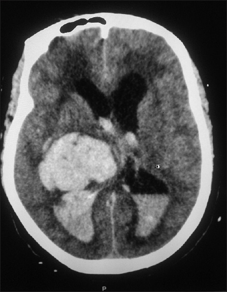
Results: There were 148 patients with complete data who were clinically diagnosed with stroke. From cranial CT findings, 56.1% of patients studied had cerebral infarct, 41.2% hemorrhage, and 2.7% normal findings. The gender distribution of cranial CT findings was not statistically significant (P = 0.09 for males and P = 0.07 for females). The parietal lobe was the most affected site for hemorrhage and infarcts accounting for 31.1% and 49.4%, respectively. The cerebellum was the least affected site. The two most commonly documented risk factors identified in this study were hypertension and diabetes mellitus accounting for 61.9%.
Conclusion: Cerebral infarct was the most common computed tomographic finding among patients with stroke, and the parietal lobe was the most common location for infarct and hemorrhage. Hypertension was a major risk factor for stroke. CT is an important imaging modality for diagnosis, differentiating infarct from hemorrhage in stroke management.
Downloads
Article Details
Section
How to Cite
References
1. Lloyd‑Jones D, Adams R, Carnethon M, De Simone G, Ferguson TB, Flegal K, et al. Heart disease and stroke statistics‑2009 update: A report
from the American Heart Association Statistics Committee and Stroke Statistics Subcommittee. Circulation 2009;119:480‑6.
2. Piliszek A, Witkowski G, Sklinda K, Szary C, Ryglewicz D, Dorobek M, et al. Comprehensive imaging of stroke – Looking for the gold standard. Neurol Neurochir Pol 2016;50:241‑50.
3. Donkor ES. Stroke in the 21 Century: A snapshot of the burden, epidemiology, and quality of life. Stroke Res Treat 2018;2018:110.
4. Wahab KW. The burden of stroke in Nigeria. Int J Stroke 2008;3:290‑2.
5. DeLaPaz RL, Wippold FJ 2nd, Cornelius RS, Amin‑Hanjani S, Angtuaco EJ, Broderick DF, et al. ACR appropriateness criteria® on
cerebrovascular disease. J Am Coll Radiol 2011;8:532‑8.
6. Neuroimaging of Acute Stoke. Available from: https://www.uptodate.com/contents/neuroimaging‑of‑acute‑ischemic‑stroke. [Last accessed on 2018 Oct 12].
7. El‑Koussy M, Schroth G, Brekenfeld C, Arnold M. Imaging of acute ischemic stroke. Eur Neurol 2014;72:309‑16.
8. Ikpeme AA, Bassey DE, Oku AO, Ephraim PE. Computerised tomography findings of cerebrovascular disease in adults Calabar, Nigeria. West Afr J Radiol 2014;21:12‑6.
9. Watila MM, Nyandaiti YW, Ibrahim A, Balarabe SA, Gezawa ID, Bakki B, et al. Risk factor profile among black stroke patients in Northeastern Nigeria. J Neurosci Behav Health 2012;4:50‑8.
10. Eze C, OkaroA, Ohagwu C. Pattern of computed tomography findings in cerebrovascular accident patients in south eastern Nigeria – A
retrospective study of 480 patients. Eur J Sci Res 2009;34:104‑9.
11. Kumar LT, Gore VN, Patil GC. The role of computed tomography in the evaluation of cerebrovascular accidents. Int J Res Med Sci 2016; 4:4305‑9.
12. Oyinloye O, Nzeh D, Adesiyun O, Ibrahim M, Akande H, Sanya E. Neuroimaging of young adults with stroke in Ilorin Nigeria. Ann Afr
Med 2015;14:82‑8.
13. Garba HY, Sule AS, Sadisu MM, Muhammad D. Pattern of computerized tomography of the brain findings in stroke patients. Ann Afr Med 2014;13:217‑20.
14. Obajimi MO, Nyame PK, Jumah KB, Wiredu EK. Computed tomographic patterns of intracranial infarcts in Ghanaians. West Afr
J Med 2002;21:121‑3.
15. Daffue K, Joubert G, Otto S. Computed tomography stroke findings and population demographics at Pelonomi hospital, Bloemfontein.
S Afr J Rad 2016;20:993.