

Diagnostic Reference Levels for Computed Tomography of the Head in Anambra State of Nigeria
Article Sidebar
Views | PDF/EPUB Downloads:
179
/ 18
/ 15
Main Article Content
Abstract
Background: Diagnostic reference levels (DRLs) were first conceptualized in 1996 by the International Commission on Radiological Protection as a result of wide variations in patient dose levels for the same examination. Current works on computed tomography (CT) doses in Nigeria produced significant variations. These observed variations, coupled with unavailable national or regional DRLs have presented the need for the establishment of standards through a dose survey.
Objective: The aim of this study is to establish DRLs for CT of the head in adult populations of Anambra State of Nigeria.
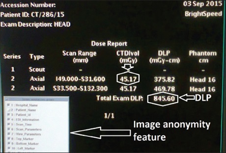
Materials and Methods: The retrospective survey was carried out from February to June 2016 in the four busiest CT centers. The digital CT population considered was those of subjects examined in 2015, and who were aged ≥18 years. Two hundred folders, comprising fifty from each center were included. The on‑screen volume computed tomography dose index (CTDIvol) and dose-length product (DLP) for the subjects were recorded. The 75th percentile was then calculated for each center to establish center-specific DRLs. Finally, a combined 75th percentile of the CTDIvol and DLP for all centers was calculated to establish the DRLs for the state. Data were analyzed using SPSS version 20.0 (SPSS Incorporated, Chicago, Illinois, USA).
Results: The digital folders of 104male and 96 female subjects with age range of 18–93 years were analyzed. The specific 75th percentile of the CTDIvol and the DLP of the centers ranged from 46 to 86 milligray (mGy) and 794 to 1785 mGy centimeters(mGy-cm), respectively. The DRLs for the State are 66 mGy and 1444 mGy-cm, respectively.
Conclusion: The DRLs for head CT in Anambra State has been derived. Although the CTDIvol is comparable to the recommendations of the European Commission, the DLP is significantly higher. Further training on dose optimization may help to bring the radiation dose in the locality at par with foreign values.
Downloads
Article Details
Section

This work is licensed under a Creative Commons Attribution-NonCommercial 4.0 International License.
This is an open access journal, and articles are distributed under the terms of the Creative Commons Attribution-NonCommercial-ShareAlike 4.0 License, which allows others to remix, tweak, and build upon the work non-commercially, as long as appropriate credit is given and the new creations are licensed under the identical terms.
How to Cite
References
1. Radiological protection and safety in medicine. A report of the International Commission on Radiological Protection. Ann ICRP
1996;26:1‑47.
2. Wall BF, Shrimpton PC. The historical development of reference doses in diagnostic radiology. Radiat Prot Dosimetry 1998;80:15‑9.
3. Foley SJ, McEntee MF, Rainford LA. Establishment of CT diagnostic reference levels in Ireland. Br J Radiol 2012;85:1390‑7.
4. Gray JE, Archer BR, Butler PF, Hobbs BB, Mettler FA Jr., Pizzutiello RJ Jr., et al. Reference values for diagnostic radiology:
Application and impact. Radiology 2005;235:354‑8.
5. Olarinoye IO, Sharifat I. A protocol for setting dose reference level for medical radiography in Nigeria: A review. Bayero J Pure Appl
Sci 2010;3:138‑41.
6. International Electrotechnical Commission. Medical Electrical Equipment‑Part 2‑44: Particular Requirements for the Safety of X‑ray Equipment for Computed Tomography. Geneva, Switzerland: IEC; 2002.
7. McCollough CH, Leng S, Yu L, Cody DD, Boone JM, McNitt‑Gray MF. CT dose index and patient dose: They are not the same thing. Radiology 2011;259:311‑6.
8. Vassileva J, RehaniMM, Al‑Dhuhli H, Al‑Naemi HM, Al‑SuwaidiJS, Appelgate K, et al. IAEA survey of pediatric CT practice in 40 countries in Asia, Europe, Latin America, and Africa: Part 1, frequency and appropriateness. AJR Am J Roentgenol 2012;198:1021‑31.
9. Martin CJ, Le Heron J, Borrás C, Sookpeng S, Ramirez G. Approaches to aspects of optimisation of protection in diagnostic radiology in six continents. J Radiol Prot 2013;33:711‑34.
10. European Commission Guidelines on Quality Criteria for Computed Tomography. Report EUR 16262 EN. Luxembourg: Office for Official Publications of the European Commission; 1999. p. 66‑78.
11. Vawda Z, Pitcher R, Akudugu J, Groenewald W. Diagnostic reference levels for paediatric computed tomography. SAfr J Radiol
2015;19:1‑4.
12. GedelAM, Gablah PG. Management of radiation dose to pediatric patients undergoing CT examination at Korle‑Bu Teaching
Hospital, Accra – Ghana. Adv Phys Theor Appl 2014;37:30‑7.
13. Wambani JS, Korir GK, Onditi EG, Korir IK. A survey of computed tomography imaging techniques and patient dose in Kenya. East
Afr Med J 2010;87:400‑7.
14. Ngaile JE, Msaki PK. Estimation of patient organ doses from CT examinations in Tanzania. J Appl Clin Med Phys 2006;7:80‑94.
15. MuhogoraWE, Ahmed NA, BeganovicA, BeniderA, Ciraj‑Bjelac O, Gershan V, et al. Patient doses in CT examinations in 18 countries: Initial results from International Atomic Energy Agency projects. Radiat Prot Dosimetry 2009;136:118‑26.
16. Ogbole G, Obed R. Radiation doses in computed tomography: Need for optimization and application of dose reference levels in
Nigeria. West Afr J Radiol 2014;21:1‑6.
17. Garba I, Engel‑Hills P, Davidson F, Tabari AM. Computed tomography dose index for head CT in northern Nigeria. Radiat Prot Dosimetry 2015;165:98‑101.
18. Abdullahi M, Shittu H, Joseph D, Aribisala A, Eshiett EP, Itopa R, et al. Diagnostic reference level for adult brain computed
tomography scans: A case study of a tertiary health care center in Nigeria. IOSR J Dent Med Sci 2015;14:66‑75.
19. Osei EK, Darko J. A survey of organ equivalent and effective doses from diagnostic radiology procedures. ISRN Radiol 2012; 2013:204346.
20. Tonkopi E, Abdolell M, Duffy S. Establishment of CT diagnostic reference levels in province Nova Scotia. Med Phys 2015; 42:32‑49.
21. Saravanakumar A, Vaideki K, Govindarajan KN, Jayakumar S. Establishment of diagnostic reference levels in computed
tomography for select procedures in Pudhuchery, India. J Med Phys 2014;39:50‑5.
22. Tsai HY, Tung CJ, Yu CC, Tyan YS. Survey of computed tomography scanners in Taiwan: Dose descriptors, dose guidance levels, and effective doses. Med Phys 2007;34:1234‑43.
23. Brix G, Nagel HD, Stamm G, Veit R, Lechel U, Griebel J, et al. Radiation exposure in multi‑slice versus single‑slice spiral CT:
Results of a nationwide survey. Eur Radiol 2003;13:1979‑91.
24. Sistrom CL. The ACR appropriateness criteria: Translation to practice and research. J Am Coll Radiol 2005;2:61‑7.
25. Tipnis S, Thampy R, Rumboldt Z, Spampinato M, Matheus G, Huda W. Radiation intensity (CTDIvol) and visibility of anatomical
structures in head CT examinations. J Appl Clin Med Phys 2016;17:5701.
26. Goo HW, Suh DS. The influences of tube voltage and scan direction on combined tube current modulation: A phantom study. Pediatr Radiol 2006;36:833‑40.
27. Aweda MA, Arogundade RA. Patient dose reduction methods in computerized tomography procedures: A review. Int J Phys Sci
2007;2:1‑9.
28. Kopp AF, Heuschmid M, Claussen CD. Multidetector helical CT of the liver for tumor detection and characterization. Eur Radiol
2002;12:745‑52.
29. Wildberger JE, Mahnken AH, Schmitz‑Rode T, Flohr T, Stargardt A, Haage P, et al. Individually adapted examination protocols for reduction of radiation exposure in chest CT. Invest Radiol 2001;36:604‑11.