

Further observations on the “spaghetti sign” in upper urinary tract hemorrhage
Article Sidebar
Views | PDF/EPUB Downloads:
22
/ 3
/ 3
Main Article Content
Abstract
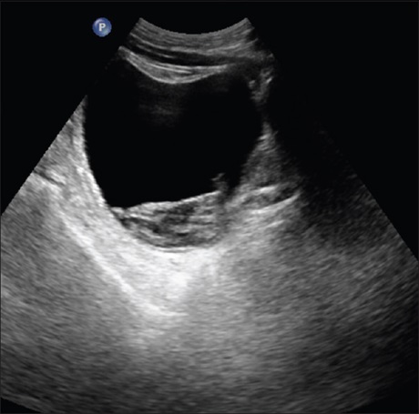
The “spaghetti sign” is recognized as a radiological sign of upper urinary tract hemorrhage. The sign was first described in the urinary bladder during intravenous urography, but it has subsequently been described on retrograde pyelography and in the urographic phase of contrast CT. We report the observation of the “spaghetti sign” in the bladder on ultrasonography and on Magnetic Resonance Urography (MRU), modalities in which the sign has not been previously described. We suggest that the observations may provide a useful guide when ultrasonography and/or MRU are employed in the search for the source of massive hematuria. We also report two additional cases of hematuria in whom the “spaghetti sign” is demonstrated in the urographic phase of contrast CT.
Downloads
Article Details
Section

This work is licensed under a Creative Commons Attribution-NonCommercial-ShareAlike 4.0 International License.
This is an open access journal, and articles are distributed under the terms of the Creative Commons Attribution-NonCommercial-ShareAlike 4.0 License, which allows others to remix, tweak, and build upon the work non-commercially, as long as appropriate credit is given and the new creations are licensed under the identical terms.
How to Cite
References
1. Komolafe F. The “spaghetti sign”: An uncommon radiologic sign of upper urinary tract hemorrhage. AJR Am J Roentgenol 1981;137:1062.
2. Dyer RB, Chen MY, Zagoria RJ. Classic signs in uroradiology. Radiographics 2004;24 Suppl 1:S247‑80.
3. Dyer RB, DiSantis DJ. The spaghetti sign. Abdom Radiol (NY) 2017;42:969‑70.
4. Komolafe F, Dahniya MH. The spaghetti sign. In: A Teaching Atlas of Case Studies in Diagnostic Imaging. New Delhi: Jaypee Publishers; 2015. p. 215‑7.
5. Eisenberg RL. The “spaghetti sign”. In: Atlas of Signs in Radiology. Ch. 2. Philadelphia: J.B. Lippincot Co.; 1985. p. 169.
6. O’Connor OJ, Fitzgerald E, Maher MM. Imaging of hematuria. AJR Am J Roentgenol 2010;195:W263‑7.
7. Frakopoulou C, Rosario DJ. Haematuria. Surgery (Oxford) 2013;31:509‑15.
8. Koehler PR, Kyaw MM. Hematuria. Med Clin North Am 1975;59:201‑32.
9. O’Connor OJ, McSweeney SE, Maher MM. Imaging of hematuria. Radiol Clin North Am 2008;46:113‑32, vii.
10. Moloney F, Murphy KP, Twomey M, O’Connor OJ, Maher MM. Haematuria: An imaging guide. Adv Urol 2014;2014:414125.
11. Leyendecker JR, Barnes CE, Zagoria RJ. MR urography: Techniques and clinical applications. Radiographics 2008;28:23‑46.
12. Brogdon BG, Silbiger ML, Colston JA Jr. Cystitis glandularis. Radiology 1965;85:470‑3.
13. Navarro JE, Huggins TJ. Cystitis glandularis: An unusual cause of ureteral obstruction. Urol Radiol 1984;6:27‑9.
14. Kauzlaric D, Barmier E, Campana A. Diagnosis of cystitis glandularis. Urol Radiol 1988;9:50‑2. [Doi: 10.1007/BF02932630].
15. Yi X, Lu H, Wu Y, Shen Y, Meng Q, Cheng J, et al. Cystitis glandularis: A controversial premalignant lesion. Oncol Lett 2014;8:1662‑4.
16. Dubbins PA, Kurtz AB, Darby J, Goldberg BB. Ureteric jet effect: The echographic appearance of urine entering the bladder. A means of identifying the bladder trigone and assessing ureteral function. Radiology 1981;140:513‑5.
17. Burge HJ, Middleton WD, McClennan BL, Hildebolt CF. Ureteral jets in healthy subjects and in patients with unilateral ureteral calculi:
Comparison with color Doppler US. Radiology 1991;180:437‑42.
18. Wu CC. Ureteric Jet. J Med Ultrasound 2010;18:141‑6.