

Role and spectrum of imaging in ovarian torsion
Article Sidebar
Views | PDF/EPUB Downloads:
342
/ 74
/ 38
Main Article Content
Abstract
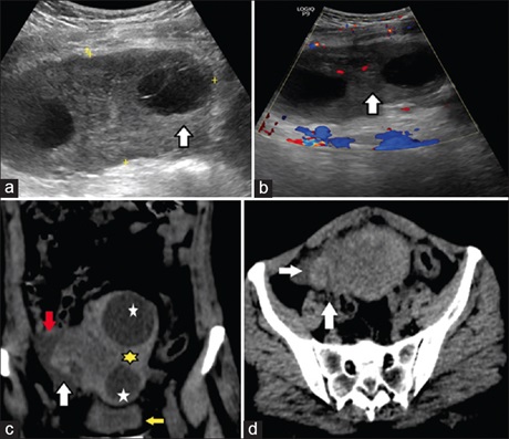
Ovarian torsion, an emergency abdominal and gynecological condition requiring immediate surgical intervention, is characterized by the twisting of ovary and its ligamentous attachment over its pedicle. As no specific clinical signs are there for accurate diagnosis, a radiologist may be the first person to make the diagnosis. Varying radiological findings on different modalities, namely ultrasonography (USG), computed tomography (CT), and magnetic resonance imaging are there characterizing ovarian torsion. Knowledge and understanding of these features can help radiologists make accurate diagnosis helping clinician for timely intervention. We here present a series of five different cases of ovarian torsion, demonstrating different and multiple imaging features of ovarian torsion on USG and CT.
Downloads
Article Details
Section
This is an open access journal, and articles are distributed under the terms of the Creative Commons Attribution-NonCommercial-ShareAlike 4.0 License, which allows others to remix, tweak, and build upon the work non-commercially, as long as appropriate credit is given and the new creations are licensed under the identical terms.
How to Cite
References
Mashiach R, Melamed N, Gilad N, Ben Shitrit G, Meizner I. Sonographic diagnosis of ovarian torsion: Accuracy and predictive factors. J Ultrasound Med 2011;30:1205‑10.
Feng JL, Zheng J, Lei T, Xu YJ, Pang H, Xie HN. Comparison of ovarian torsion between pregnant and non‑pregnant women at reproductive ages: Sonographic and pathological findings. Quant Imaging Med Surg 2020;10:137‑47.
Wattar B, Rimmer M, Rogozinska E, Macmillian M, Khan KS, Al Wattar BH. Accuracy of imaging modalities for adnexal torsion: A systematic review and meta‑analysis. BJOG 2021;128:37‑44.
Dawood MT, Naik M, Bharwani N, Sudderuddin SA, Rockall AG, Stewart VR. Adnexal torsion: Review of radiologic appearances. Radiographics 2021;41:609‑24.
Swenson DW, Lourenco AP, Beaudoin FL, Grand DJ, Killelea AG, McGregor AJ. Ovarian torsion: Case‑control study comparing the sensitivity and specificity of ultrasonography and computed tomography for diagnosis in the emergency department. Eur J Radiol 2014;83:733‑8.
Dhanda S, Quek ST, Ting MY, Rong CYH, Ting EYS, Jagmohan P, et al. CT features in surgically proven cases of ovarian torsion—a pictorial review. Br J Radiol 2017;90:20170052.
Duigenan S, Oliva E, Lee SI. Ovarian torsion: Diagnostic features on CT and MRI with pathologic correlation. AJR Am J Roentgenol 2012;198:W122‑31.
Singh T, Prabhakar N, Singla V, Bagga R, Khandelwal N. Spectrum of magnetic resonance imaging findings in ovarian torsion. Pol J Radiol 2018;83:e588‑99.
Khalil R, El‑Dieb L. Sonographic and MRI Features of Ovarian Torsion. Egypt J Radiol Nucl Med 2016; 47:621‑9.
10. Huang C, Hong MK, Ding DC. A review of ovary torsion. Ci Ji Yi Xue Za Zhi 2017;29:143‑7.
11. Ghulmiyyah L, Nassar A, Sassine D, Khoury S, Nassif J, Ramadan H, et al. Accuracy of Pelvic Ultrasound in Diagnosing Adnexal Torsion. Radiology research and practice 2019;2019:1406291. doi: 10.1155/2019/1406291.
12. Hiller N, Appelbaum L, Simanovsky N, Lev Sagi A, Aharoni D, Sella T. CT features of adnexal torsion. AJR Am J Roentgenol 2007;189:124‑9.
13. Tonolini M, Foti PV, Costanzo V, Mammino L, Palmucci S, Cianci A, et al. Cross‑sectional imaging of acute gynaecologic disorders: CT and
MRI findings with differential diagnosis‑part I: Corpus luteum and haemorrhagic ovarian cysts, genital causes of haemoperitoneum and
adnexal torsion. Insights Imaging 2019;10:119.
14. Higashide R, Tsukada T, Ichikawa M, Sakamoto M, Shimabukuro K. Ovarian torsion due to ovarian hyperstimulation syndrome diagnosed
by sonographic whirlpool sign in the first trimester of pregnancy: A case report. Radiol Case Rep 2023;18:3386‑9. 15.
Krishnan S, Kaur H, Bali J, Rao K. Ovarian torsion in infertility management – Missing the diagnosis means losing the ovary: A high price to pay. J Hum Reprod Sci 2011;4:39‑42.
16. Gomes MM, Cavalcanti LS, Reis RL, Silva EJ, Dutra JB, de Melo Leite AF. Twist and shout: Magnetic resonance imaging findings in ovarian torsion. Radiol Bras 2019;52:397‑402.
17. Singh S, Sasmal PK, Nagarajan K. CT imaging in predicting ovarian torsion: Report of two cases, with and without infarction. Cureus
2021;13:e17082.
18. Jung SI, Park HS, Yim Y, Jeon HJ, Yu MH, Kim YJ, et al. Added value of using a CT coronal reformation to diagnose adnexal torsion. Korean
J Radiol 2015;16:835‑45.
19. Rha SE, Byun JY, Jung SE, Jung JI, Choi BG, Kim BS, et al. CT and MR imaging features of adnexal torsion. Radiographics 2002;22:283‑94.
20. Franco PN, García Baizán A, Aymerich M, Maino C, Frade Santos S, Ippolito D, et al. Gynaecological causes of acute pelvic pain: Common and not‑so‑common imaging findings. Life (Basel) 2023;13:2025.
21. Lee JH, Park SB, Shin SH, Jang JC, Lee WC, Jeong AK, et al. Value of intra‑adnexal and extra‑adnexal computed tomographic imaging
features diagnosing torsion of adnexal tumor. J Comput Assist Tomogr 2009;33:872‑6.
22. Lee MS, Moon MH, Woo H, Sung CK, Oh S, Jeon HW, et al. CT findings of adnexal torsion: A matched case‑control study. PLoS One 2018;13:e0200190.
23. Ghossain MA, Hachem K, Aoun NJ, Haddad Zebouni S, Mansour F, Suidan JS, et al. Spontaneous detorsion of the ovary: Can it be diagnosed by MRI? J Magn Reson Imaging 2006;24:880‑5