

A Case of Status Epilepticus: A Giant Panda Dropped the Hint
Article Sidebar
Views | PDF/EPUB Downloads:
201
/ 24
/ 30
Main Article Content
Abstract
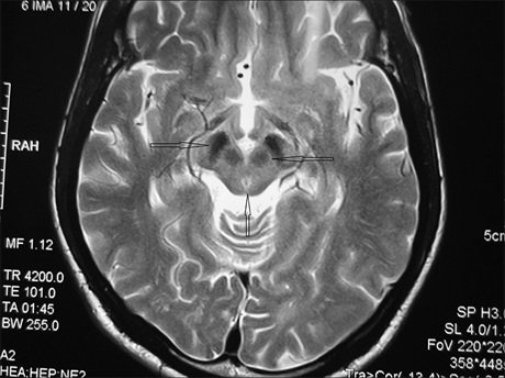
Wilson's disease can manifest as neurological disorder without hepatic involvement. However, first presentation as status epilepticus is extremely rare. Herein, we report a case where a 21-year-old male presented with status epilepticus. Clinical background, biochemical tests and typical magnetic resonance imaging findings in the form of “Face of giant Panda sign” confirmed the diagnosis of Wilson's disease.
Downloads
Article Details
Section

This work is licensed under a Creative Commons Attribution-NonCommercial 4.0 International License.
This is an open access journal, and articles are distributed under the terms of the Creative Commons Attribution-NonCommercial-ShareAlike 4.0 License, which allows others to remix, tweak, and build upon the work non-commercially, as long as appropriate credit is given and the new creations are licensed under the identical terms.
How to Cite
References
1. Dening TR, Berrios GE, Walshe JM. Wilson’s disease and epilepsy. Brain 1988;111:1139‑55.
2. Shukla R, Desai P, Vinod P. Wilson’s disease presenting as status epilepticus. J Assoc Physicians India 2006;54:887‑9.
3. Peters RA, Shorthouse M, Walshe JM. The effect of Cu+2 on the membrane ATPase and its relationship to initiation of convulsions.
J Physiol 1965;181:27‑8.
4. Ulkii T, KadirA, Recep A. Status epilepticusin a case of Wilson’s disease during D‑penicillamine treatment. Swiss Med Wkly 2003; 103:446‑7.
5. Benbir G, Gunduz A, Ertan S, Ozkara C. Partial status epilepticus induced by hypocupremia in a patient with Wilson’s disease.
Seizure 2010;19:602‑4.
6. Sinha S, Taly AB, Ravishankar S, Prashanth LK, Venugopal KS, Arunodaya GR, et al. Wilson’s disease: Cranial MRI observations
and clinical correlation. Neuroradiology 2006;48:613‑21.
7. Kim YE, Yun JY, Yang HJ, Kim HJ, Jeon BS. Unusual epileptic deterioration and extensive white matter lesion during treatment in Wilson’s disease. BMC Neurol 2013 25;13:127.
8. Hitoshi S, Iwata M, Yoshikawa K. Mid‑brain pathology of Wilson’s disease: MRI analysis of three cases. J Neurol Neurosurg Psychiatry 1991;54:624‑6.
9. Rutledge JN, Hilal SK, Silver AJ, Defendini R, Fahn S. Study of movement disorders and brain iron by MR. Am J Roentgenol 1987; 149:365‑79.
10. Prashanth LK, Sinha S, Taly AB, Vasudev MK. Do MRI features distinguish Wilson’s disease from other early onset extrapyramidal
disorders? An analysis of 100 cases. Mov Disord 2010;25:672‑8.