

Herlyn‑Werner‑Wunderlich syndrome: A rare cause of pain in the left iliac fossa
Article Sidebar
Views | PDF/EPUB Downloads:
14
/ 2
/ 2
Main Article Content
Abstract
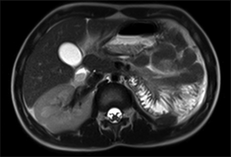
Alterations of the Müllerian ducts are rare but often treatable causes of infertility. Uterus didelphys is caused by a complete or almost complete lack of fusion of the Müllerian ducts during embryological development. As a result, two separate symmetrical uterine cavities develop with two cervices and no communication between these cavities; this is often associated with a vaginal septum, which can have a transverse wall that blocks one of the hemivaginas. Symptoms begin to develop in menarche, and complications related to a retrograde menstrual flow arise, along with pelvic adhesions and endometriosis. Some kidney abnormalities may occur. Magnetic resonance imaging can be used to diagnose and distinguish surgically correctable forms of Müllerian duct alterations. The surgical approach depends on the imaging findings. The objective of this article is to describe the radiological characteristics of the Herlyn-Werner-Wunderlich syndrome.
Downloads
Article Details
Section

This work is licensed under a Creative Commons Attribution-NonCommercial-ShareAlike 4.0 International License.
This is an open access journal, and articles are distributed under the terms of the Creative Commons Attribution-NonCommercial-ShareAlike 4.0 License, which allows others to remix, tweak, and build upon the work non-commercially, as long as appropriate credit is given and the new creations are licensed under the identical terms.
How to Cite
References
1. Moore KL, Persaud TV. The urogenital system: The development of the genital system. Clinically Oriented Embryology. 6th ed. Philadelphia,
PA: WB Saunders Company; 1998.
2. Mandava A, Prabhakar RR, Smitha S. OHVIRA syndrome (obstructed hemivagina and ipsilateral renal anomaly) with uterus didelphys, an
unusual presentation. J Pediatr Adolesc Gynecol 2012;25:e23‑5.
3. Shah DK, Laufer MR. Obstructed hemivagina and ipsilateral renal anomaly (OHVIRA) syndrome with a single uterus. Fertil Steril 2011; 96:e39‑41.
4. Guillán‑Maquieira C, Sánchez‑Merino JM, Méndez‑Díaz C. OHVIRA Sindrome (obstructed hemivagina and ipsilateral renal anomaly)
associated to uterus didelphys. Prog Obstet Ginecol 2012;55:281‑4.
5. Gholoum S, Puligandla PS, Hui T, Su W, Quiros E, Laberge JM, et al. Management and outcome of patients with combined vaginal septum,
bifid uterus, and ipsilateral renal agenesis (Herlyn‑werner‑wunderlich syndrome). J Pediatr Surg 2006;41:987‑92.
6. O’Neill MJ, Yoder IC, Connolly SA, Mueller PR. Imaging evaluation and classification of developmental anomalies of the female reproductive system with an emphasis on MR imaging. AJR Am J Roentgenol 1999;173:407‑16.
7. Osornio‑Sánchez V, Santana‑Ríos Z, Fulda‑Graue SD, Pérez‑Becerra R, Urdiales‑Ortiz A, Martínez A, et al. Herlyn Werner Wünderlich síndrome. Review of the literature and case report. Rev Mex Urol 2012;72:31‑4.
8. Broseta E, Boronat F, Ruiz JL, Alonso M, Osca JM, Jiménez‑Cruz JF, et al. Urological complications associated to uterus didelphys with
unilateral hematocolpos. A case report and review of the literature. Eur Urol 1991;20:85‑8.
9. Bermejo‑Espinosa N, Isusi F, Díaz R. Case solution: Syndrome Herlyn Werner Wünderlich. Radiologia 2010;52:366‑8.
10. Orazi C, Lucchetti MC, Schingo PM, Marchetti P, Ferro F. Herlyn‑werner‑wunderlich syndrome: Uterus didelphys, blind hemivagina and ipsilateral renal agenesis. Sonographic and MR findings in 11 cases. Pediatr Radiol 2007;37:657‑65.
11. Daniels P, Donoso M, Arraztoa JA. Hysteroscopic resection of the vaginal septum in Herlyn-Werner-Wünderlich syndrome: A case report.
Rev Chil Obstet Ginecol 2010;75:185‑8.