

Calcium caplet mimicking renal calculi
Article Sidebar
Views | PDF/EPUB Downloads:
86
/ 36
/ 33
Main Article Content
Abstract
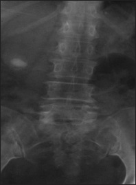
We present a 72-year-old woman, a known hypertensive patient with excruciating right upper back pain that worsened over 3 days. No fever, vomiting, hematuria, or lower urinary tract symptoms. Examination revealed right renal angle tenderness. Antero-posterior [Figure 1] lumbosacral X-ray showed an oblong radio-opaque shadow of calcific density projected over the lower pole of the right kidney opposite the third lumbar vertebra.
Other findings included straightening of the normal lumbar lordosis (seen on the lateral projection) and moderate degenerative changes of the lumbar spine. However, the pedicles, disk spaces posterior elements, and the pre-vertebral soft tissue space were normal. Abdominal ultrasound scan performed 2 h after the initial radiographs did not reveal any renal or gall bladder calculi. A repeat abdominal X-ray done 3h after the initial radiograph showed no radio-opaque shadow.
Downloads
Article Details
Section

This work is licensed under a Creative Commons Attribution-NonCommercial 4.0 International License.
This is an open access journal, and articles are distributed under the terms of the Creative Commons Attribution-NonCommercial-ShareAlike 4.0 License, which allows others to remix, tweak, and build upon the work non-commercially, as long as appropriate credit is given and the new creations are licensed under the identical terms.
How to Cite
References
1. Mshelbwala PM, Ameh EA, Mbibu HN. Urinary stone in children. Nig J Surg Res 2005;7:238‑43.
2. Stoller ML. Urinary stone disease. In: Tanagho A, McAninch JW, editors. Smith’s General Urology. 16th ed. New York City: The McGraw‑Hill Companies; 2004. p. 256‑90.
3. Portis AJ, Sundaram CP. Diagnosis and initial management of kidney stones. Am Fam Physician 2001;63:1329‑38.
4. Kaur P, Chauhan A, Singh G, Kataria S, Kalra R. Primary squamous cell carcinoma of kidney ‑ A case report and review of literature. Int J Nephrol 2010;6. Available from: http://www.ispub.com/journal/the‑internet‑journal‑of‑nephrology/volume‑6‑number‑1/primary‑squamous‑cell‑carcinoma‑of‑kidney‑a‑case‑report‑and‑review‑of‑literature.html [Last accessed on 2011 Jul 13].
5. Wein AJ. Surgical anatomy of the retroperitoneum, adrenals, kidneys, and ureters. In: Kavoussi LR, Novick AC, Partin AW, Peters CA, editors. Campbell‑Walsh Urology. 9th ed. New York: Saunders Elsevier; 2007. p. 3‑37.
6. Kambadakone AR, Eisner BH, Catalano OA, Sahani DV. New and evolving concepts in the imaging and management of urolithiasis: Urologists’ perspective. Radiographics 2010;30:603‑23.
7. Ha M, MacDonald RD. Impact of CT scan in patients with first episode of suspected nephrolithiasis. J Emerg Med 2004;27:225‑31.
8. Schwartz G, Lipschitz S, Becker JA. Detection of renal calculi: The value of tomography. AJR Am J Roentgenol 1984;143:143‑5.