

Pharyngeal Dimensions in Skeletal Class I, II, and III Orthodontic Patients in a Nigerian Population
Article Sidebar
Views | PDF/EPUB Downloads:
360
/ 55
/ 49
Main Article Content
Abstract
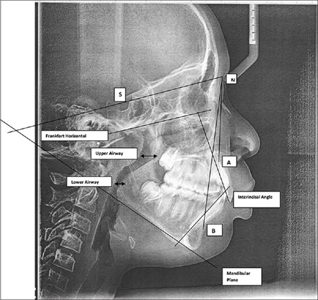
Background: Routine lateral cephalometric radiographs can determine upper and lower pharyngeal airway constriction or patency
depending on the dentofacial skeletal discrepancy. Appropriate orthodontic treatment that would maintain or improve the airway
patency can be considered if the width of the airway in the various skeletal classes is determined.
Aim: The aim of this study was to evaluate the upper and lower pharyngeal widths in skeletal Class I, II, and III untreated orthodontic patients in Benin City, Nigeria.
Materials and Methods: In this study, 188 lateral cephalometric radiographs comprising three groups based on the ANB angle:
Class I (ANB 2–4°), Class II (ANB >4), and Class III (ANB < 2) were analyzed using the method described by McNamara. The
vertical facial pattern (the Sella–Nasion‑GoGn angle) and palatal length were also determined. The differences between groups
and correlations between variables were determined with the Students t‑test and the Spearman correlation coefficient, respectively.
Results: The mean upper and lower pharyngeal width for skeletal Classes I were 10.56 ± 3.67 mm and 11.14 ± 3.79 mm, respectively.
Skeletal Class II had the narrowest upper airway width, whereas skeletal Class III had the narrowest lower airway widths, respectively.
The palatal length was 9.04 mm in males and 8.6 mm in females, and there was a highly statistically significant difference P < 0.05
between hyperdivergent facial pattern and the upper pharyngeal width. There was a significant difference between skeletal pattern II
and the upper pharyngeal width.
Conclusion: Pharyngeal dimensions should be taken into consideration when managing patients with skeletal patterns II and III and the hyperdivergent facial patterns.
Downloads
Article Details
Section

This work is licensed under a Creative Commons Attribution-NonCommercial 4.0 International License.
This is an open access journal, and articles are distributed under the terms of the Creative Commons Attribution-NonCommercial-ShareAlike 4.0 License, which allows others to remix, tweak, and build upon the work non-commercially, as long as appropriate credit is given and the new creations are licensed under the identical terms.
How to Cite
References
1. Jacobson A, Jacobson RL. Radiographic Cephalometry from Basics to 3‑D Imaging. 2nd ed. Chicago: Quintessence Publishing Co.; 2006.
2. McNamara JA Jr. A method of cephalometric evaluation. Am J Orthod 1984;86:449‑69.
3. Posnick JC. Orthognathic Surgery. Principles and Practice. Vol. 1, Ch. 10. Philadelphia: Elseiver Saunders Publishing Co.; 2014. p. 287‑97.
4. Aloufi F, Preston CB, Zawawi KH. Changes in the upper and lower pharyngeal airway spaces associated with rapid maxillary expansion. ISRN Dent 2012;2012:290964.
5. Jia P, Fu M, Zeng X. Changes of upper airway morphology induced by mandibular advancement in patients with obstructive sleep apnea syndrome. Beijing Da Xue Xue Bao 2003;35:663‑7.
6. Gonçalves Rde C, Raveli DB, Pinto Ados S. Effects of age and gender on upper airway, lower airway and upper lip growth. Braz Oral Res 2011;25:241‑7.
7. Bishara SE, Jakobsen JR, Hession TJ, Treder JE. Soft tissue profile changes from 5 to 45 years of age. Am J Orthod Dentofacial Orthop 1998; 114:698‑706.
8. Memon S, Fida M, Shaikh A. Comparison of different craniofacial patterns with pharyngeal widths. J Coll Physicians Surg Pak 2012;22:302‑6.
9. Batool I, Shaheed M, Rizvi SA, Abbas A. Comparison of upper and lower pharyngeal airway space in class II high and low angle cases. Pak Oral Dent J 2010;30:81‑4.
10. Isiekwe MC, Sowemimo GO. Cephalometric findings in a normal Nigerian population sample and adult Nigerians with unrepaired clefts. Cleft Palate J 1984;21:323‑8.
11. Utomi IL. Cephalometric norms in Nigerian Hausa‑fulanis. West Afr J Med 2004;23:119‑22.
12. Ajayi EO. Cephalometric norms of Nigerian children. Am J Orthod Dentofacial Orthop 2005;128:653‑6.
13. Ifesanya JU. An update on cephalometrics among Nigerians: Ascertaining prevalent jaw patterns. Br J Med Med Res 2014;4:3092‑100.
14. Adesina BA, Otuyemi OD, Kolawole KA, Adeyemi AT. Two‑dimensional analysis of oro‑pharyngeal airway space area in patients with bimaxillary protrusion. West Afr J Orthod 2013;2:5‑11.
15. Houston WJ. The analysis of errors in orthodontic measurements. Am J Orthod 1983;83:382‑90.
16. Bruntz LQ, Palomo JM, Baden S, Hans MG. A comparison of scanned lateral cephalograms with corresponding original radiographs. Am J Orthod Dentofacial Orthop 2006;130:340‑8.
17. Dobrowolska‑Zarzycka M, Dunin‑Wilczynska I, Mitura I, Dabala M. Evaluation of upper airways depth among patients with skeletal class I and III. Folia Morphol (Warsz) 2013;72:155‑60.
18. Kirjavainen M, Kirjavainen T. Upper airway dimensions in class II malocclusion. Effects of headgear treatment. Angle Orthod 2007; 77:1046‑53.
19. Eslamian L, Badiee MR, Yousefinia S, Kharazifard MJ. Radiographic assessment of upper airway size in skeletal sagittal and vertical jaw discrepancies. J Islam Dent Assoc 2014;26:15‑20.
20. Gupta S, Subrahmanya RM. Assessment of oropharyngeal widths in individuals with different facial skeleton patterns. Nitte Univ J Health Sci 2014;4:34‑8.
21. Stellzig‑Eisenhauer A, Meyer‑Marcotty P. Interaction between otorhinolaryngology and orthodontics: Correlation between the nasopharyngeal airway and the craniofacial complex. Laryngorhinootologie 2010;89 Suppl 1:S72‑8.
22. Sparks RJ, Ngan P, Martin C, Razmus T, Mah J, Gunel E. A Comparison of Airway Dimensions Among Different Skeletal Craniofacial Patterns. New York: Nova Science Publishers Inc; 2012. p. 401‑26.
23. Tourné LP. Growth of the pharynx and its physiologic implications. Am J Orthod Dentofacial Orthop 1991;99:129‑39.
24. Batool I, Shaheed M, Rizvi AS, Abbas A. Comparison of upper and lower pharyngeal airway space in class II high and low angle cases. Pak Oral Dent J 2010;30:81‑4.
25. de Freitas MR, Alcazar NM, Janson G, de Freitas KM, Henriques JF. Upper and lower pharyngeal airways in subjects with class I and class II malocclusions and different growth patterns. Am J Orthod Dentofacial Orthop 2006;130:742‑5.
26. Gu M, McGrath CP, Wong RW, Hägg U, Yang Y. Cephalometric norms for the upper airway of 12‑year‑old Chinese children. Head Face Med 2014;10:38.
27. Riquelme A, Green LJ. Palatal width, height, and length in human twins. Angle Orthod 1970;40:71‑9.