

Radiological And Clinical Pattern Of Pleural Effusion In Ilorin.
Article Sidebar
Views | PDF/EPUB Downloads:
190
/ 16
Main Article Content
Abstract
Background: Pleural effusion (PE) is the commonest manifestation of pleural disease and may herald pathologies from other parts of the body. Etiology varies with age and geographical location. Chest radiography is an essential component of early assessment though there are suggestions to apply chest ultrasonography also early in patient evaluation. This study aims to determine the radiological and clinical pattern of pleural effusions based on clinical and radiological diagnosis of pleural effusion and to correlate this with etiology.
Method: A retrospective analysis of 276 plain Chest X-rays (CXR) of patients diagnosed clinically to have PE over a period of 6% years in the University of Teaching Hospital was conducted.
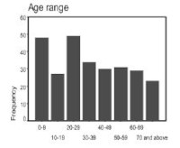
Result: A bimodal age distribution involving the first and second decades was demonstrated. Etiological factors were identified in 95.6% of cases. Chronic inflammation and pyogenic effusion accounted for 47.6%. Amongst children, adolescents and young adults, the percentage rose to 62.0%. Heart failure was responsible for PE in 18.1% of all the cases and 63.3% of patients -50years. More than half of PE occurred in the right hemithorax (53.1%). Pyogenic and malignant effusion showed predilection for the right side (75.6% and 58.3% respectively). Heart failure accounted for 57.1% of all patients with bilateral effusions.
Conclusion: We found that chronic inflammation and pyogenic effusion accounted for a greater proportion of etiological factors. Conventional CXR is still valuable as a first line investigative modality.
Downloads
Article Details
Section

This work is licensed under a Creative Commons Attribution-NonCommercial 4.0 International License.
This is an open access journal, and articles are distributed under the terms of the Creative Commons Attribution-NonCommercial-ShareAlike 4.0 License, which allows others to remix, tweak, and build upon the work non-commercially, as long as appropriate credit is given and the new creations are licensed under the identical terms.
How to Cite
References
1. HT Mocelin, GB Fischer. Epidemiology, presentation and treatment of pleural effusion, Paed. Resp. Reviews. 2002; 3: 292-297
2. GW Saton Jr, RH Ingram Jr. Disorders of the pleurs, hila and mediastinum: pleural effusion. ACP medicine online. 2002; ©2002 WebMD Inc.
3. VV Garrido, JF Sancho, HB Blasco et al. Diagnosis and treatment of pleural effusion. Recommendation of the Spanish Society of Pulmonology and Thoracic Surgery (SEPAR). Arch. Bronconrumol. 2006; 42(7): 349-72
4. RS Andrade, MM Maddaus. Pleural effusion: Approach to patient with pleural effusion. ACS Surgery online. 2002; 2002 WebMD. Inc.
5. J Ferrer, J Roldán. Clinical management of the patient with pleural effusion. Eur J Radiol. 2000; 34:76-86
6. NM Rahman, FV Gleeson. directions in the treatment of infected pleural effusion. Clinical radiology. 2006; 61:719-722
7. K Kocijancic, M Tercelj, K Vidmar, et al. The value of inspiratory-expiratory lateral decubitus views in the diagnosis of small pleural effusions. Clinical Radiol. 1999; 54: 595-597.
8. A Quadri, AH Thomson. Pleural fluid associated with chest infection. Paed. Resp. Reviews, 2002; 3; 349-355.
9. L Valdes, A Pose, E San Jose, et al. Tuberculous pleural effusion. Eur. J. Int. Med. 2003; 14:77-88
10. DR Quintero, LL Fan. Approach to pleural effusions and empyemas. Pard. Resp. Reviews 2004; 5(Suppl A): S151-8152
11. Caksen H, Ozturk MK, Yuksel S et al. Parapneumonic pleural effusion and empyema in childhood. J Emergency Med. 2003 May, 24 (4): 474-476