

Apparent diffusion coefficient; is it an effective index for differentiating between types of lung cancer brain metastases?
Article Sidebar
Views | PDF/EPUB Downloads:
515
/ 97
/ 52
Main Article Content
Abstract
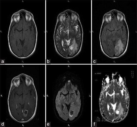
Background: The apparent diffusion coefficient (ADC) values of tumors are highly correlated with tumor cellularity and used as a neuroimaging marker with the potential to differentiate between major histological subtypes. Here, we will attempt to determine the sensitivity and specificity of the ADC to distinguish between types of metastatic brain metastases from lung cancer.
Methods: One hundred and fifty‑six patients (136 [%87, 18] male, 20 [%12.82] female) admitted to our hospital with the diagnosis of primary lung cancer were included in the study. In addition to conventional magnetic resonance imaging sequences, Diffusion-weighted imaging (DWI) and ADC images were evaluated qualitatively and quantitatively.
Results: We found hyperintensity in most of the metastatic lesions on a qualitatively evaluated DWI sequence. In quantitative assessment according to ADC value comparisons between the different histologic subtype metastatic lung carcinoma groups, small-cell carcinoma (SCLC) had the highest value (1.93 × 10‒3mm2/s ± 0.95) and nonsmall‑cell‑combined (NSCCLC) type was the least (0.55 × 10‒3 mm2/s ± 0.46). When we tried to distinguish lung cancer-induced brain metastases into two main groups as SCC and NSC by considering the mean ADC ratios we obtained 0.65 ± 0.14 for SCC and 1.51± 0.30 for NSC. On the other hand, there was no significant statistical difference between the specific histological subtype groups with comparison of ADC values (P > 0.05).
Conclusion: Quantitatively quantified DWI-ADC can distinguish metastatic lesions from the normal brainparenchyma. Although we realized whether differentiation of SCLC and non-SCLC in brain metastases can be achieved with DWI, we could not define any correlation between DWI/ADC values and primary histology of the metastatic foci. We believe that more accurate results can be achieved with advanced studies with more patients included and common sequence features.
Downloads
Article Details
Section
This is an open access journal, and articles are distributed under the terms of the Creative Commons Attribution-NonCommercial-ShareAlike 4.0 License, which allows others to remix, tweak, and build upon the work non-commercially, as long as appropriate credit is given and the new creations are licensed under the identical terms.
How to Cite
References
1. Duygulu G, Ovali GY, Calli C, Kitis O, Yünten N, Akalin T, et al. Intracerebral metastasis showing restricted diffusion: Correlation with
histopathologic findings. Eur J Radiol 2010;74:117‑20.
2. Hayashida Y, Hirai T, Morishita S, Kitajima M, Murakami R, Korogi Y, et al. Diffusion‑weighted imaging of metastatic brain tumors: Comparison with histologic type and tumor cellularity. AJNR Am J Neuroradiol 2006;27:1419‑25.
3. Liu K, Ma Z, Feng L. Apparent diffusion coefficient as an effective index for the therapeutic efficiency of brain chemoradiotherapy for brain metastases from lung cancer. BMC Med Imaging 2018;18:30.
4. Dirier A, Karadayi B. Comparison of two different radiotherapy schedules in lung cancer patients with brain metastasis. Dicle Med J 2006;33:134‑8.
5. Chi A, Komaki R. Treatment of brain metastasis from lung cancer. Cancers (Basel) 2010;2:2100‑37.
6. Pellerino A, Internò V, Muscolino E, Mo F, Bruno F, Pronello E. et al. Leptomeningeal metastases from non‑small cell lung cancer: State of
the art and recent advances. J Cancer Metastasis Treat 2020;6:41.
7. Demir OI, Obuz F, Sağol O, Dicle O. Contribution of diffusion‑weighted MRI to the differential diagnosis of hepatic masses. Diagn Interv
Radiol 2007;13:81‑6.
8. van Everdingen KJ, van der Grond J, Kappelle LJ, Ramos LM, Mali WP. Diffusion‑weighted magnetic resonance ımaging in acute stroke. Stroke
1998;29:1783‑90.
9. Back T, Hoehn‑Berlage M, Kohno K, Hossmann KA. Diffusion nuclear magnetic resonance imaging in experimental stroke. Correlation with cerebral metabolites. Stroke 1994;25:494‑500.
10. Le Bihan D, Turner R, Douek P, Patronas N. Diffusion MR imaging: Clinical applications. AJR Am J Roentgenol 1992;159:591‑9.
11. Yoshikawa T, Kawamitsu H, Mitchell DG, Ohno Y, Ku Y, Seo Y, et al. ADC measurement of abdominal organs and lesions using parallel
imaging technique. AJR Am J Roentgenol 2006;187:1521‑30.
12. Ichikawa T, Haradome H, Hachiya J, Nitatori T, Araki T. Diffusion‑weighted MR imaging with a single‑shot echoplanar sequence: Detection and characterization of focal hepatic lesions. AJR Am J Roentgenol 1998;170:397‑402.
13. Calli C, Kitis O, Yunten N, Yurtseven T, Islekel S, Akalin T. Perfusion and diffusion MR imaging in enhancing malignant cerebral tumors. Eur J Radiol 2006;58:394‑403.
14. Vollmer RT. The effect of cell size on the pathologic diagnosis of small and large cell carcinomas of the lung. Cancer 1982;50:1380‑3.
15. Dietrich O, Biffar A, Reiser MF, Baur‑Melnyk A. Diffusion‑weighted imaging of bone marrow. Semin Musculoskelet Radiol 2009;13:134‑44.
16. Berghoff AS, Spanberger T, Ilhan‑Mutlu A, Magerle M, Hutterer M, Woehrer A, et al. Preoperative diffusion‑weighted imaging of single
brain metastases correlates with patient survival times. PLoS One 2013;8:e55464.
17. Tang G, Liu Y, Li W, Yao J, Li B, Li P. Optimization of b value in diffusion‑weighted MRI for the differential diagnosis of benign and malignant vertebral fractures. Skeletal Radiol 2007;36:1035‑41.
18. Caravan I, Ciortea CA, Contis A, Lebovici A. Diagnostic value of apparent diffusion coefficient in differentiating between high‑grade gliomas and brain metastases. Acta Radiol 2018;59:599‑605.
19. Karaarslan E, Arslan A. Diffusion weighted MR imaging in non‑infarct lesions of the brain. Eur J Radiol 2008;65:402‑16.
20. Yurdakul AS, Calısır HC, Demirag F, Taci N, Ogretensoy M. The distribution of hystological types of lung cancer (Analysis of 2216 cases). Turk Thorac J 2002;3:59‑65.
21. Tas SG, Ak H, Ceylan E, Yaycıoglu S, Meteoglu I, Cıldag O. Asymptomatic solitary pons metastasis in small cell lung cancer: A case report. ADU Med Fac J 2009;10:41‑3.
22. Zakaria R, Das K, Radon M, Bhojak M, Rudland PR, Sluming V, et al. Diffusion‑weighted MRI characteristics of the cerebral metastasis to brain boundary predicts patient outcomes. BMC Med Imaging 2014;14:26.
23. Müller SJ, Khadhraoui E, Neef NE, Riedel CH, Ernst M. Differentiation of brain metastases from small and non‑small lung cancers using apparent diffusion coefficient (ADC) maps. BMC Med Imaging 2021;21:70.
24. Meyer HJ, Fiedler E, Kornhuber M, Spielmann RP, Surov A. Comparison of diffusion‑weighted imaging findings in brain metastases of different origin. Clin Imaging 2015;39:965‑9.
25. Tien RD, Felsberg GJ, Friedman H, Brown M, MacFall J. MR imaging of high‑grade cerebral gliomas: Value of diffusion‑weighted echoplanar pulse sequences. AJR Am J Roentgenol 1994;162:671‑7.
26. Brunberg JA, Chenevert TL, McKeever PE, Ross DA, Junck LR, Muraszko KM, et al. In vivo MR determination of water diffusion coefficients and diffusion anisotropy: Correlation with structural alteration in gliomas of the cerebral hemispheres. AJNR Am J Neuroradiol 1995;16:361‑71.
27. Shaefer PW, Grant PE, Gonzalez RG. Diffusion weighted MR imaging of the brain. Radiology 2000;217:331‑45.
28. Han C, Huang S, Guo J, Zhuang X, Han H. Use of a high b‑value for diffusion weighted imaging of peritumoral regions to differentiate
high‑grade gliomas and solitary metastases. J Magn Reson Imaging 2015;42:80‑6.