

Morphometric Evaluation of Soft Palate in Oral Submucous Fibrosis‑A Digital Cephalometric Analysis
Article Sidebar
Views | PDF/EPUB Downloads:
209
/ 64
/ 39
Main Article Content
Abstract
Aims: The present clinico-radiological study was done to evaluate the morphological variants of soft palate in oral submucous fibrosis (OSMF) patients using digital lateral cephalometry. Different variations in the morphology of soft palate were compared with stages of OSMF. Further, soft palate morphology in OSMF patients was compared radiographically with that of normal population.
Materials and Methods: A total number of 100 patients who were a part of this study were divided in two equal Groups. Group 1 comprised of 50 patients clinically diagnosed with OSMF and Group 2 included 50 routine patients.
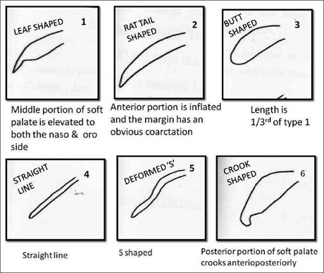
Results: Six different morphological variants of soft palate were found. Among the study Groups, type 1soft palate was most commonly seen (56%) whereas type 5 was the least common variant. Majority of patients belonged to stage II OSMF and type 1soft palate was commonly seen in this stage of disease whereas butt shaped soft palate (type 3) was more common in stage III OSMF.
Conclusion: In OSMF, type 1 and 2 are commonly seen but as the diseases advances, these are replaced by type 3 and 6 variants. In OSMF patients, there in reduction in the anterio-posterior dimension of soft palate.
Downloads
Article Details
Section

This work is licensed under a Creative Commons Attribution-NonCommercial 4.0 International License.
This is an open access journal, and articles are distributed under the terms of the Creative Commons Attribution-NonCommercial-ShareAlike 4.0 License, which allows others to remix, tweak, and build upon the work non-commercially, as long as appropriate credit is given and the new creations are licensed under the identical terms.
How to Cite
References
1. Rao AB. Idiopathic palatal fibrosis. Br J Surg 1962;50:23‑5.
2. Wahi PN, Luthra UK, Kapur VL. Submucus fibrosis of oral cavity. Histomorphological studies. Br J Cancer 1966;20:676‑87.
3. More CB, Gupta S, Joshi J, Varma SN. Classification system for oral submucous fibrosis. J Indian Acad Oral Med Radiol 2012;1:24‑9.
4. You M, Li X, Wang H, Zhang J, Wu H, Liu Y, et al. Morphological variety of the soft palate in normal individuals: A digital
cephalometric study. Dentomaxillofac Radiol 2008;37:344‑9.
5. Kumar DK, Gopal KS. Morphological variants of soft palate in normal individuals: A digital cephalometric study. J Clin Diagn
Res 2011;5:1310‑3.
6. Bacon WH, Turlot JC, Krieger J, Stierle JL. Cephalometric evaluation of pharyngeal obstructive factors in patients with sleep
apneas syndrome. Angle Orthod 1990;60:115‑22.
7. Lyberg T, Krogstad O, Djupesland G. Cephalometric analysis in patients with sleep apnea syndrome: II. Soft tissue morphology.
J Laryngol Otol 1989;103:293‑7.
8. Pepin JL, Ferretti G, Veale D, Romand P, Coulomb M, Brambilla C, et al. Somnofluroscopy, computed tomography and cephalometry in the assessment of the airway in obstructive sleep apnea. Thorax 1992;47:150‑6.