

Artificial intelligence and machine learning in neurosurgery: A review of diagnostic significance and treatment planning efficiency
Article Sidebar
Views | PDF/EPUB Downloads:
402
/ 132
/ 62
Main Article Content
Abstract
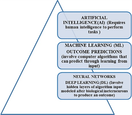
This review analyzes the significance of artificial intelligence (AI) and deep learning (DL) approaches used in radiology in neurosurgery patients and compares AI applications with human models to determine the applicability of AI in disease diagnosis, decision-making, and outcome prediction. A systematic review was conducted from 1997 to 2020 from the PubMed (MEDLINE) database. The search strategy adhered to guidelines outlined by the Preferred Reporting Items for Systematic Reviews and Meta-Analyses. The keywords used for the literature search included “Deep learning,” “Neurosurgery,” “Artificial Intelligence,” “Brain,” “Magnetic resonance imaging-MRI Brain,” and “Machine learning.” The studies focusing on the significance of DL and comparing AI applications with radiologists or clinical experts to enhance diagnostic protocols were included, whereas non-English articles, animal studies, articles lacking full text, and publications such as commentaries, technical notes, abstracts, editorials, opinions, and letters were excluded. A total of 24 articles were included in the review. The P value was observed in 44 out of 63 outcome measures (70%), out of which in 26 out of 63 outturn measures, artificial application subset machine learning (ML) has a significant edge over clinical diagnosis (P < 0.05). The review highlights the potential impact of AI-driven advancements in clinical radiology on enhancing treatment plans for neurosurgery patients, emphasizing the benefits of early intervention, cost reduction, time-saving approaches, and judicious health-care resource utilization. The study’s limitations include potential constraints in identifying relevant literature due to the selected search scope and inclusion criteria, not including studies published outside the specified timeframe and database, and a small number of included studies. Consequently, there is a risk of overlooking innovative methodologies or ground-breaking studies contributing to a more comprehensive understanding of AI applications in neurosurgery. Furthermore, the exclusion of certain publication types, such as commentaries, and conference papers may limit the diversity of different perspectives. However, the study highlights the potential of ML in neurosurgery and the importance of addressing variability in study design, patient populations, and outcome measures in future research to enhance the applicability of AI-driven approaches in clinical practice. It is imperative to recognize and address these challenges to understand the opportunities and limitations inherent in the integration of AI in neurosurgical practice.
Downloads
Article Details
Section
This is an open access journal, and articles are distributed under the terms of the Creative Commons Attribution-NonCommercial-ShareAlike 4.0 License, which allows others to remix, tweak, and build upon the work non-commercially, as long as appropriate credit is given and the new creations are licensed under the identical terms.
How to Cite
References
1. Rudie JD, Rauschecker AM, Bryan RN, Davatzikos C, Mohan S. Emerging applications of artificial intelligence in neuro‑oncology. Radiology 2019;290:607‑18.
2. Panch T, Szolovits P, Atun R. Artificial intelligence, machine learning and health systems. J Glob Health 2018;8:020303.
3. YaoAD, ChengDL, Pan I, Kitamura F. Deep learning in neuroradiology: A systematic review of current algorithms and approaches for the new
wave of imaging technology. Radiol Artif Intell 2020;2:e190026.
4. LeCun Y, Bengio Y, Hinton G. Deep learning. Nature 2015;521:436‑44.
5. Nucci CG, De Bonis P, Mangiola A, Santini P, Sciandrone M, Risi A, et al. Intracranial pressure wave morphological classification: Automated
analysis and clinical validation. Acta Neurochir (Wien) 2016;158:581‑.
6. Swinburne NC, Schefflein J, Sakai Y, Oermann EK, Titano JJ, Chen I, et al. Machine learning for semi‑automated classification of glioblastoma, brain metastasis and central nervous system lymphoma using magnetic resonance advanced imaging. Ann Transl Med 2019;7:232.
7. Titano JJ, Badgeley M, Schefflein J, Pain M, Su A, Cai M, et al.Automated deep‑neural‑network surveillance of cranial images for acute neurologic events. Nat Med 2018;24:1337‑41.
8. Topol EJ. High‑performance medicine: The convergence of human and artificial intelligence. Nat Med 2019;25:44‑56.
9. Kitajima M, Hirai T, Katsuragawa S, Okuda T, Fukuoka H, Sasao A,et al. Differentiation of common large sellar‑suprasellar masses effect of artificial neural network on radiologists’ diagnosis performance. Acad Radiol 2009;16:313‑20.
10. Yamashita K, Yoshiura T, Arimura H, Mihara F, Noguchi T, Hiwatashi A, et al. Performance evaluation of radiologists with artificial neural network for differential diagnosis of intra‑axial cerebral tumors on MR images. AJNR Am J Neuroradiol 2008;29:1153‑8.
11. Bidiwala S, Pittman T. Neural network classification of pediatric posterior fossa tumors using clinical and imaging data. Pediatr Neurosurg 2004;40:8‑15.
12. Arle JE, Perrine K, Devinsky O, Doyle WK. Neural network analysis of preoperative variables and outcome in epilepsy surgery. J Neurosurg 1999;90:998‑1004.
13. Awuah WA, Adebusoye FT, Wellington J, David L, Salam A, Weng Yee AL, et al. Recent outcomes and challenges of artificial intelligence, machine learning, and deep learning in neurosurgery. World Neurosurg×2024;23:100301.
14. Buchlak QD, Esmaili N, Leveque JC, Bennett C, Farrokhi F, Piccardi M. Machine learning applications to neuroimaging for glioma detection and classification: An artificial intelligence augmented systematic review. J Clin Neurosci 2021;89:177‑98.
15. Buchlak QD, Esmaili N, Leveque JC, Farrokhi F, Bennett C, Piccardi M,et al. Machine learning applications to clinical decision support in neurosurgery: An artificial intelligence augmented systematic review. Neurosurg Rev 2020;43:1235‑53.
16. Mofatteh M. Neurosurgery and artificial intelligence. AIMS Neurosci 2021;8:477‑95.
17. Groiss SJ, Wojtecki L, Südmeyer M, Schnitzler A. Deep brain stimulation in Parkinson’s disease. Ther Adv Neurol Disord 2009;2:20‑8.
18. Pilaz LJ, Liu J, Joshi K, Tsunekawa Y, Musso CM, D’Arcy BR, et al.Subcellular mRNA localization and local translation of Arhgap11a in radial glial progenitors regulates cortical development. Neuron 2023;111:839‑56.e5.
19. Mofatteh M. Neurodegeneration and axonal mRNA transportation. Am J Neurodegener Dis 2021;10:1‑12.
20. Juntu J, Sijbers J, De Backer S, Rajan J, Van Dyck D. Machine learning study of several classifiers trained with texture analysis features to differentiate benign from malignant soft‑tissue tumors in T1‑MRI images. J Magn Reson Imaging 2010;31:680‑9.
21. Zhao ZX, Lan K, Xiao JH, Zhang Y, Xu P, Jia L, et al. A new method to classify pathologic grades of astrocytomas based on magnetic resonance imaging appearances. Neurol India 2010;58:685‑90.
22. Emblem KE, Nedregaard B, Hald JK, Nome T, Due Tonnessen P, Bjornerud A. Automatic glioma characterization from dynamic susceptibility contrast imaging: Brain tumor segmentation using knowledge‑based fuzzy clustering. J Magn Reson Imaging 2009;30:1‑10.
23. Abdolmaleki P, Mihara F, Masuda K, Buadu LD. Neural networks analysis of astrocytic gliomas from MRI appearances. Cancer Lett 1997;118:69‑78.
24. Sinha M, Kennedy CS, Ramundo ML. Artificial neural network predicts CT scan abnormalities in pediatric patients with closed head injury. J Trauma 2001;50:308‑12.
25. Cohen KB, Glass B, Greiner HM, Holland Bouley K, Standridge S, Arya R, et al. Methodological issues in predicting pediatric epilepsy surgery candidates through natural language processing and machine learning. Biomed Inform Insights 2016;8:11‑8.
26. Cuocolo R, Ugga L, Solari D, Corvino S, D’Amico A, Russo D, et al.Prediction of pituitary adenoma surgical consistency: Radiomic data mining and machine learning on T2‑weighted MRI. Neuroradiology 2020;62:1649‑56.
27. Dolz J, Betrouni N, Quidet M, Kharroubi D, Leroy HA, Reyns N, et al.Stacking denoising auto‑encoders in a deep network to segment the brainstem on MRI in brain cancer patients: A clinical study. Comput Med Imaging Graph 2016;52:8‑18.
28. Hashido T, Saito S, Ishida T. A radiomics‑based comparative study on arterial spin labeling and dynamic susceptibility contrast perfusion‑weighted imaging in gliomas. Sci Rep 2020;10:6121.
29. Clarke LP, Velthuizen RP, Clark M, Gaviria J, Hall L, Goldgof D,et al. MRI measurement of brain tumor response: Comparison of visual metric and automatic segmentation. Magn Reson Imaging 1998;16:271‑9.
30. Chiang S, Levin HS, Haneef Z. Computer‑automated focus lateralization of temporal lobe epilepsy using fMRI. J Magn Reson Imaging 2015;41:1689‑94.
31. Kassahun Y, Perrone R, De Momi E, Berghöfer E, Tassi L, Canevini MP, et al. Automatic classification of epilepsy types using ontology‑based and genetics‑based machine learning. Artif Intell Med 2014;61:79‑88.
32. Kerr WT, Nguyen ST, Cho AY, Lau EP, Silverman DH, Douglas PK,et al. Computer‑aided diagnosis and localization of lateralized temporal lobe epilepsy using interictal FDG‑PET. Front Neurol 2013;4:31.
33. Lee JS, Lee DS, Kim SK, Lee SK, Chung JK, Lee MC, et al. Localization of epileptogenic zones in F‑18 FDG brain PET of patients with temporal lobe epilepsy using artificial neural network. IEEE Trans Med Imaging 2000;19:347‑55.
34. Ledley RS, Lusted LB. Reasoning foundations of medical diagnosis; symbolic logic, probability, and value theory aid our understanding of how physicians reason. Science 1959;130:9‑21.
35. Lodwick GS, Keats TE, Dorst JP. The coding of roentgen images for computer analysis as applied to lung cancer. Radiology 1963;81:185‑200.
36. Rughani AI, Dumont TM, Lu Z, Bongard J, Horgan MA, Penar PL, et al. Use of an artificial neural network to predict head injury outcome.
J Neurosurg 2010;113:585‑90.
37. Haug PJ. Uses of diagnostic expert systems in clinical care. Proc Annu Symp Comput Appl Med Care 1993;1993:379‑83.
38. Emblem KE, Pinho MC, Zöllner FG, Due Tonnessen P, Hald JK, Schad LR, et al. A generic support vector machine model for preoperative glioma survival associations. Radiology 2015;275:228‑34.
39. Ambinder EP. A history of the shift toward full computerization of medicine. J Oncol Pract 2005;1:54‑6.
40. Ganapathy N, Swaminathan R, Deserno TM. Deep learning on 1‑D biosignals: A taxonomy‑based survey. Yearb Med Inform 2018;27:98‑109.
41. Kuhlmann L, Lehnertz K, Richardson MP, SchelterB, ZaveriHP. Seizure prediction – Ready for a new era. Nat Rev Neurol 2018;14:618‑30.
42. Kwon JM, Lee Y, Lee Y, Lee S, Park J. An algorithm based on deep learning for predicting in‑hospital cardiac arrest. J Am Heart Assoc 2018;7:e008678.
43. Shin HC, Roth HR, Gao M, Lu L, Xu Z, Nogues I, et al. Deep convolutional neural networks for computer‑aided detection: CNN architectures, dataset characteristics and transfer learning. IEEE Trans Med Imaging 2016;35:1285‑98.
44. Kermany DS, Goldbaum M, Cai W, Valentim CC, Liang H, Baxter SL,et al. Identifying medical diagnoses and treatable diseases by image‑based deep learning. Cell 2018;172:1122‑31.e9.
45. Katzman JL, Shaham U, Cloninger A, Bates J, Jiang T, Kluger Y. DeepSurv: Personalized treatment recommender system using a Cox proportional hazards deep neural network. BMC Med Res Methodol 2018;18:24.
46. Jiménez J, Škalič M, Martínez Rosell G, De Fabritiis G. K (DEEP): Protein‑ligand absolute binding affinity prediction via 3D‑convolutional neural networks. J Chem Inf Model 2018;58:287‑96.
47. Kalinin AA, Higgins GA, Reamaroon N, Soroushmehr S, Allyn Feuer A, Dinov ID, et al. Deep learning in pharmacogenomics: From gene regulation to patient stratification. Pharmacogenomics 2018;19:629‑50.
48. Jiang S, Chin KS, Tsui KL. A universal deep learning approach for modeling the flow of patients under different severities. Comput Methods Programs Biomed 2018;154:191‑203.
49. Vranas KC, Jopling JK, Sweeney TE, Ramsey MC, Milstein AS, Slatore CG, et al. Identifying Distinct Subgroups of ICU patients: A machine learning approach. Crit Care Med 2017;45:1607‑15.
50. Rajkomar A, Oren E, Chen K, DaiAM, Hajaj N, Hardt M, et al. Scalable and accurate deep learning with electronic health records. NPJ Digit Med 2018;1:18.
51. Shariff S, Kantawala B, Mkrtchyan A, Mirzaei F, Grigoryan V. Artificial intelligence in neurosurgery: Pre, intra and post operation applications.
J Surg 2023;8:1895.
52. Kozel G, Gurses ME, Gecici NN, Gökalp E, Bahadir S, Merenzon MA,et al. Chat‑GPT on brain tumors: An examination of artificial intelligence/machine learning’s ability to provide diagnoses and treatment plans for example neuro‑oncology cases. Clin Neurol Neurosurg 2024;239:108238.
53. Tangsrivimol JA, Schonfeld E, Zhang M, Veeravagu A, Smith TR, Härtl R, et al. Artificial intelligence in neurosurgery: A state‑of‑the‑art review from past to future. Diagnostics (Basel) 2023;13:2429.
54. Noh SH, Cho PG, Kim KN, Kim SH, Shin DA. Artificial intelligence for neurosurgery: Current state and future directions. J Korean Neurosurg Soc 2023;66:113‑20.
55. Charles YP, Lamas V, Ntilikina Y. Artificial intelligence and treatment algorithms in spine surgery. Orthop Traumatol Surg Res 2023;109:103456.
56. Yagi M, Yamanouchi K, Fujita N, Funao H, Ebata S. Revolutionizing spinal care: Current applications and future directions of artificial intelligence and machine learning. J Clin Med 2023;12:4188.
57. Iqbal J, Jahangir K, Mashkoor Y, Sultana N, Mehmood D, Ashraf M,et al. The future of artificial intelligence in neurosurgery: A narrative
review. Surg Neurol Int 2022;13:536.
58. González CastroV, Valdés Hernández MD, Chappell FM, Armitage PA, Makin S, Wardlaw JM. Reliability of an automatic classifier for brain enlarged perivascular spaces burden and comparison with human performance. Clin Sci (Lond) 2017;131:1465‑81.