

Age‑related ultrasonographic mammary gland density patterns: Implication for breast cancer risk
Article Sidebar
Views | PDF/EPUB Downloads:
453
/ 86
/ 31
Main Article Content
Abstract
Introduction: Mammary gland/breast density is important because it is a known biomarker for breast cancer risk. However, the sensitivity of mammography decreases with high breast density found in younger age group. Ultrasound is considered as the first-line examination in the classification of breast density and in the detection and characterization of breast lesions. This study aims to evaluate the relationship between age and ultrasonographic breast density pattern and its implication for breast cancer risk.
Materials and Methods: This was a community-based cross-sectional, exploratory, descriptive study involving 658 females. Breast ultrasonographic scans were performed using a Sonoace X1 Machine with a 7.5 MHz transducer. The lesions detected and classified by ultrasonography as benign or malignant were further subjected to cytopathology.
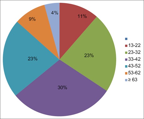
Results: Modal age group of the participants ranged from 33 to 43 years representing 29.8%. There was significant correlation (P<0.01) between ultrasonographic mammary gland density pattern and age, the age group of <33 years demonstrated predominant fibroglandular density pattern with mostly benign lesions, while the age group of 33–53 years demonstrated predominant heterogeneous breast density pattern with most of the malignant lesions in this age group, making it the high-risk group for breast cancer.
Conclusion: The relationship between age and ultrasonographic breast density is inversely proportional and not absolute. It also concludes that ultrasonography is a reliable screening tool in the diagnostic process for mammary gland lesions, and as an imaging tool, it is the preferred modality in dense breast. The heterogeneous fibroglandular pattern emerged as the high-risk group for breast cancer, especially in middle age.
Downloads
Article Details
Section
This is an open access journal, and articles are distributed under the terms of the Creative Commons Attribution-NonCommercial-ShareAlike 4.0 License, which allows others to remix, tweak, and build upon the work non-commercially, as long as appropriate credit is given and the new creations are licensed under the identical terms.
How to Cite
References
1. Wolf JN. Breast parenchyma and their changes with age. Radiology 1976;121:545‑52.
2. Bland KI, CopelandIIIV EM, Klimberg S. Anatomy of the breast, axilla, chest wall, and related metastatic sites. In: The Breast; Comprehensive Management of Benign and Malignant Diseases. 5th ed. Elsevier Inc; 2018. p. 20‑36.
3. Adetifa FA, Ojikutu RK. Prevalence and trends in breast cancer in Lagos State, Nigeria. Afr Res Rev 2009;3:01‑15.
4. WHO. Early detection. Cancer control: New guide to cancer and early diagnosis. Elsevier Inc.: World Health Organization; 2017.
5. Obajimi MO, Adeniji‑Sofoluwe AT, Oluwasola AO, Adedokun BO, Mosuro OA, Adeoye AO, et al. Screening mammography in Ibadan:
Our experience. Niger J Basic Clin Sci 2015;12:74‑80.
6. Geisel J, Raghu M, Hooley R. The role of ultrasound in breast cancer screening: The case for and against ultrasound. Semin Ultrasound CT MR 2018;39:25‑34.
7. Mendelson EB, Berg WA, Merritt CR. Toward a standardized breast ultrasound lexicon, BI‑RADS: Ultrasound. Semin Roentgenol 2001; 36:217‑25.
8. Berg WA. Rationale for a trial of screening breast ultrasound: American College of Radiology Imaging Network (ACRIN) 6666. AJR Am J
Roentgenol 2003;180:1225‑8.
9. Jemal A, Bray F, Center MM, Ferlay J, Ward E, Forman D. Global cancer statistics. CA Cancer J Clin 2011;61:69‑90.
10. Rapelyea JA, Marks CG. Breast Imaging: Breast Ultrasound Past, Present, and Future. Editor, Kuzmiak CM, Publisher: Intech Open,
2017. DOI: 10.5772/intechopen.69790.
11. Huay‑Ben P. The role of ultrasound in early cancer detection. J Med Ultrasound 2016;24:138‑41.
12. O’Connell AM. The many roles of ultrasound in breast malignancy. Appl Radiol 2009;38:1‑6.
13. Boyd N, Berman H, Zhu J, Martin LJ, Yaffe MJ, Chavez S, et al. The origins of breast cancer associated with mammographic density:
A testable biological hypothesis. Breast Cancer Res 2018;20:17.
14. Checka CM, Chun JE, Schnabel FR, Lee J, Toth H. The relationship of mammographic density and age: Implications for breast cancer
screening. AJR Am J Roentgenol 2012;198:W292‑5.
15. Gundry KR. Breast ultrasound: Indications and findings. Clin Obstet Gynecol 2016;59:380‑93.
16. American College of Radiology Breast imaging reporting and data system. BI‑RADS atlas. In: American College of Radiology BI‑RADS
Atlas. American College of Radiology; 2013.
17. Bukhari MH, Arshad M, Jamal S, Niazi S, Bashir S, Bakhshi IM, et al. Use of fine‑needle aspiration in the evaluation of breast lumps. Patholog Res Int 2011;2011:689521.
18. Jakes RW, Duffy SW, Ng FC, Gao F, Ng EH. Mammographic parenchymal patterns and risk of breast cancer at and after a prevalence
screen in Singaporean women. Int J Epidemiol 2000;29:11‑9.
19. Akande HJ, Olafimihan BB, Oyinloye OI. Mammographic parenchymal patterns in asymptomatic women. Saudi J Med Med Sci 2017;5:232‑7.
20. Obajimi MO, Adeniji‑Sofoluwe AT, Adedokun BO, Soyemi TO, Bassey OS. Ultrasonographic breast pattern in females in Ibadan,
Nigeria. Ann Afr Med 2014;13:145‑50.
21. D’Orsi CJ, Sickles EA, Mendelson EB, Morris EA. Breast Imaging Reporting and Data System: ACR BI‑RADS – Breast Imaging Atlas. 5th ed., Vol. 272. Reston: American College of Radiology; 2013. p. 2‑15.
22. Nwaneri A, Osuala EO, Okpala PU, Emesowum AC, Iheanacho P. Knowledge and awareness of breast cancer among rural females in
Umuowa Orlu local government area Imo State, South East, Nigeria. Niger J Clin Pract 2017;20:489‑94.
23. Olarinoye‑Akorede SA, Adamu A, Balogun MS. Sonographic breast density pattern among Nigerian women in Zaria. Niger J Basic Clin
Sci 2018;2:138‑41.
24. Pike MC, Krailo MD, Henderson BE, Casagrande JT, Hoel DG. ‘Hormonal’ risk factors, ‘breast tissue age’ and the age‑incidence of
breast cancer. Nature 1983;303:767‑70.
25. Maskarinec G, Pagano I, Lurie G, Kolonel LN. A longitudinal investigation of mammographic density: The multiethnic cohort. Cancer Epidemiol Biomarkers Prev 2006;15:732‑9.
26. Ginsburg OM, Martin LJ, Boyd NF. Mammographic density, lobular involution, and risk of breast cancer. Br J Cancer 2008;99:1369‑74.