

Normal Pediatric Lumbar Lordosis: Measurement of Magnitude and Age of Maximum Development Using Three Radiographic Techniques
Article Sidebar
Views | PDF/EPUB Downloads:
289
/ 62
/ 46
Main Article Content
Abstract
Background: A retrospective measurement of lumbar lordosis (LL) in normal supine lateral lumbosacral spine radiographs of 27
children aged 0.04–14.00 years. Measurement of the LL may aid in the early diagnosis and management of some pediatric conditions
before irreversible neurologic change occurs. They include spondylolisthesis (congenital or acquired); achondroplasia and muscular
dystrophies are less common. The sagittal radiographic lumbar lordotic angle is poorly documented in normal pediatric population.
Objective: To evaluate the magnitude and age of maximum development of the normal pediatric LL using three different radiographic
techniques.
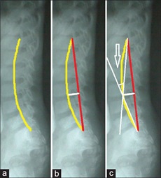
Methods: Ferguson (for lumbosacral angle [LSA]), Cobb (for Cobb angle) and tangential radiologic assessment of LL (for TRALL angle) were the methods used. Data were analyzed with SPSS statistics version 20.0 (Chicago, IL, USA). P < 0.05 was considered significant.
Results: LSA varied from 15° to 62°, Cobb angle 15–65° and TRALL angle 20–46°. The mean (standard deviation) of LSA, Cobb, and TRALL angles were 35.8 (10.3)°, 35.6 (13.7)°, and 32.3 (7.3),° respectively; the 0.95 confidence interval for the LSA was 27.6–44.5°, Cobb angle 27.2–50.7°, and TRALL angle 26.8–40.1°. Each angle showed no significant gender difference. The major part of estimated adult LL was gained during the first 5 years of life; the second peak occurred in the 11–14 years age‑group.
Conclusion: In children under 15 years, poor management of pathologies affecting LL can cause irreversible neurologic damage
arising from spinal deformity.
Downloads
Article Details
Section

This work is licensed under a Creative Commons Attribution-NonCommercial 4.0 International License.
This is an open access journal, and articles are distributed under the terms of the Creative Commons Attribution-NonCommercial-ShareAlike 4.0 License, which allows others to remix, tweak, and build upon the work non-commercially, as long as appropriate credit is given and the new creations are licensed under the identical terms.
How to Cite
References
1. Gelb DE, Lenke LG, Bridwell KH, Blanke K, McEnery KW. An analysis of sagittal spinal alignment in 100 asymptomatic middle and older aged volunteers. Spine (Phila Pa 1976) 1995;20:1351‑8.
2. Bogduk N, Twomey LT. Clinical Anatomy of the Lumbar Spine. 2nd ed. New York: Churchill Livingstone; 1991. p. 45‑7.
3. O’Rahilly R, Muller F, Meyer DB. The human vertebral column at the end of the embryonic period proper 1. The column as a whole. J Anat 1980;131(Pt 3):565‑75.
4. Bagnall KM, Harris PF, Jones PR. A radiographic study of the human fetal spine 1. The development of the secondary cervical curvature. J Anat 1977;123(Pt 3):777‑82.
5. Panattoni GL, Todros T. Postural aspects of the human fetal spine. Morphometric and functional study. Panminerva Med 1988;30:250‑3.
6. Voutsinas SA, MacEwen GD. Sagittal profiles of the spine. Clin Orthop Relat Res 1986;210:235‑42.
7. Oliver J, Middleditch A. Lumbar spine. In: Functional Anatomy of the Spine. Oxford: Butterworth‑Heinemann; 1998. p. 36‑58.
8. Norkin CC, White DJ. Joint Motion: Method of Measuring and Recording. Chicago: American Academy of Orthopaedic Surgeons; 1965. p. 48‑9.
9. Burdett RG, Brown KE, Fall MP. Reliability and validity of four instruments for measuring lumbar spine and pelvic positions. Phys Ther 1986;66:677‑84.
10. Ferguson AB. Clinical and roentgen interpretation of lumbosacral spine. Radiology 1934;22:548‑58.
11. Ferguson AB. Roentgen Diagnosis of the Extremities and Spine. 2nd ed. New York: Paul B. Hoeber, Inc.; 1949. p. 382‑3.
12. Cobb JR. Outline for the study of scoliosis. In: Thomson JE, Blount WP, editors. American Academy of Orthopaedic Surgeons, Instructional Course Lectures. Vol. 5. Ann Arbor: JW Edwards; 1948. p. 261‑75.
13. Troyanovich SJ, Harrison DE, Harrison DD, Holland B, Janik TJ. Further analysis of the reliability of the posterior tangent lateral lumbar radiographic mensuration procedure: Concurrent validity of computer‑aided X‑ray digitization. J Manipulative Physiol Ther 1998;21:460‑7.
14. Chernukha KV, Daffner RH, Reigel DH. Lumbar lordosis measurement. A new method versus Cobb technique. Spine (Phila Pa 1976) 1998; 23:74‑9.
15. Chen YL. Vertebral centroid measurement of lumbar lordosis compared with the Cobb technique. Spine (Phila Pa 1976) 1999;24:1786‑90.
16. Hong JY, Suh SW, Modi HN, Hur CY, Song HR, Park JH. Reliability analysis for radiographic measures of lumbar lordosis in adult scoliosis: A case‑control study comparing 6 methods. Eur Spine J 2010;19:1551‑7.
17. de Oliveira TS, Candotti CT, La Torre M, Pelinson PP, Furlanetto TS, Kutchak FM, et al. Validity and reproducibility of the measurements
obtained using the flexicurve instrument to evaluate the angles of thoracic and lumbar curvatures of the spine in the sagittal plane. Rehabil Res Pract 2012;2012:186156.
18. Hart DL, Rose SJ. Reliability of a noninvasive method for measuring the lumbar curve*. J Orthop Sports Phys Ther 1986;8:180‑4.
19. Rajabi R, Seidi F, Mohamadi F. Which method is accurate when using the flexible ruler to measure the lumbar curvature angle? Deep pint or midpoint of arch? World Appl Sci J 2008;4:849‑52.
20. Seidi F, Rajabi R, Ebrahimi TI, Tavanai AR, Moussavi SJ. The Iranian flexible ruler reliability and validity in lumbar lordosis measurements. World J Sport Sci 2009;2:95‑9.
21. Youdas JW, Suman VJ, Garrett TR. Reliability of measurements of lumbar spine sagittal mobility obtained with the flexible curve. J Orthop Sports Phys Ther 1995;21:13‑20.
22. Babai E, Khodamoradi A, Mosavi Z, Bahari S. An innovative software method for measuring lumbar lordosis. Ann Biol Res 2012;3:204‑13.
23. López‑Miñarro PA, Muyor JM, Belmonte F, Alacid F. Acute effects of hamstring stretching on sagittal spinal curvatures and pelvic tilt. J Hum Kinet 2012;31:69‑78.
24. Willner S. Spinal pantograph – A non‑invasive technique for describing kyphosis and lordosis in the thoraco‑lumbar spine. Acta Orthop Scand 1981;52:525‑9.
25. Souza Filho JC, Abras AC, Carvalho MT, Souza MG, Souza AT, Costa LO. Analysis of the interexaminer reliability of two clinical tests to measure the flexion range of motion of the lumbar spine. Physiatric Minutes 2007;14:214‑8.
26. Macintyre NJ, Bennett L, Bonnyman AM, Stratford PW. Optimizing reliability of digital inclinometer and flexicurve ruler measures of spine curvatures in postmenopausal women with osteoporosis of the spine: An illustration of the use of generalizability theory. ISRN Rheumatol 2011;2011:571698.
27. Fernand R, Fox DE. Evaluation of lumbar lordosis. A prospective and retrospective study. Spine (Phila Pa 1976) 1985;10:799‑803.
28. Vrtovec T, Pernus F, Likar B. A review of methods for quantitative evaluation of spinal curvature. Eur Spine J 2009;18:593‑607.
29. Salisbury PJ, Porter RW. Measurement of lumbar sagittal mobility. A comparison of methods. Spine (Phila Pa 1976) 1987;12:190‑3.
30. Spiegel DA, Dormans JP. The spine. In: Kliegman RM, Behrman RE, Jenson HB, Stanton BF, editors. Nelson Textbook of Pediatrics. 19th ed., ch. 671. Philadelphia, PA: Saunders Elsevier; 2011.
31. Hansson T, Bigos S, Beecher P, Wortley M. The lumbar lordosis in acute and chronic low‑back pain. Spine (Phila Pa 1976) 1985;10:154‑5.
32. Murrie VL, Dixon AK, Hollingworth W, Wilson H, Doyle TA. Lumbar lordosis: Study of patients with and without low back pain. Clin Anat 2003; 16:144‑7.
33. Beasley M. Lumbar Lordosis; 2015. Available from: http://www.Carta.anthropogeny.org/moca/topics/lumbar‑lordosis.
[Last accessed on 2015 May 28].
34. Reichmann S, Lewin T. The development of the lumbar lordosis. A post mortem study on excised lumbar spines. Arch Orthop Unfallchir 1971;69:275‑85.
35. Knott P, Betsch M. Evaluating Spinal Deformity Using Surface Topography. SSTSG Website, Spine and Surface Topography Study Group; 2013. Available from: http://www.sstsg.org/related‑literature.html. [Last accessed on 2015 May 28].
36. Berryman F, Pynsent P, Fairbank J, Disney S. A new system for measuring three‑dimensional back shape in scoliosis. Eur Spine J 2008;17:663‑72.
37. Thometz JG, Liu XC, Lyon R, Harris GF. Variability in three‑dimensional measurements of back contour with raster stereography in normal subjects. J Pediatr Orthop 2000;20:54‑8.
38. Propst‑Proctor SL, Bleck EE. Radiographic determination of lordosis and kyphosis in normal and scoliotic children. J Pediatr Orthop 1983; 3:344‑6.
39. Mac‑Thiong JM, Berthonnaud E, Dimar JR 2nd, Betz RR, Labelle H. Sagittal alignment of the spine and pelvis during growth. Spine (Phila Pa 1976) 2004;29:1642‑7.
40. Giglio CA, Volpon JB. Development and evaluation of thoracic kyphosis and lumbar lordosis during growth. J Child Orthop 2007;1:187‑93.