

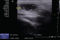
Suppurated Inguinal Node mimicking a Strangulated Inguinal Ovarian Hernia on Ultrasound
Article Sidebar
Views | PDF/EPUB Downloads:
109
/ 57
/ 26
Main Article Content
Abstract
.
Downloads
Article Details
Section
This is an open access journal, and articles are distributed under the terms of the Creative Commons Attribution-NonCommercial-ShareAlike 4.0 License, which allows others to remix, tweak, and build upon the work non-commercially, as long as appropriate credit is given and the new creations are licensed under the identical terms.
How to Cite
References
1. Park HR, Park SB, Lee ES, Park HJ. Sonographic evaluation of inguinal lesions. Clin Imaging 2016;40:949‑55.
2. Narci A, Korkmaz M, Albayrak R, Sözübir S, Güvenç BH, Köken R, et al. Preoperative sonography of nonreducible inguinal masses in girls. J Clin Ultrasound 2008;36:409‑12.
3. Jedrzejewski G, Stankiewicz A, Wieczorek AP. Uterus and ovary hernia of the canal of Nuck. Pediatr Radiol 2008;38:1257‑8.
4. Artas H, Gurbuzer N. Inguinal hernia containing both ovaries and the uterus in an infant. J Ultrasound Med 2012;31:1138‑9.
5. Aydin R, Polat AV, Ozaydin I, Aydin G. Gray‑scale and color Doppler ultrasound imaging findings of an ovarian inguinal hernia and torsion of the herniated ovary: A case report. Pediatr Emerg Care 2013;29:364‑5.