

Assessement Of Glycerol, Gelatin And Agar Gels As Equivalent Materials For Mammalian Organs In Proton Nuclear Magnetic Resonance Imaging
Article Sidebar
Views | PDF/EPUB Downloads:
100
/ 42
Main Article Content
Abstract
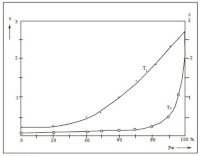
The physical NMR parameters of glycerol, gelatin and agar gels have been studied with the view of using them as materials for mimicking mammalianorgans. The spin-lattice and the spin-spin relaxation times T and T1 2 respectively for these materials with varied water contents have been measured, using the CXP-100 Brucker NMR spectrometer and the HP-9836 calculator. The results show that the ranges of T -values are 40-2521ms, 342-1 2054 ms and 942-2665 ms for glycerol, gelatin and agar gels, while the ranges for T -values are 17-1072 ms, 42-1022 ms2 and 13-433 ms respectively.
Downloads
Article Details
Section

This work is licensed under a Creative Commons Attribution-NonCommercial 4.0 International License.
This is an open access journal, and articles are distributed under the terms of the Creative Commons Attribution-NonCommercial-ShareAlike 4.0 License, which allows others to remix, tweak, and build upon the work non-commercially, as long as appropriate credit is given and the new creations are licensed under the identical terms.
How to Cite
References
1. Kuntz ID and Zipp A. “Water in Biological Systems” Physiol. In Med., 1977; Vol. 297, No 5, pp. 262-266.
2. De Certaines JD. “Measurement and Meaning of Relaxation Times Specific and Non-Specific Variation in Cancer” Annali dell' Institute Superiore di Sanita 1983; Vol. 18, No 20 pp. 107-120.
3. Bakker CJG, de Graaf CN and Van Dijk P. “Derivation of Quantitative Information in NMR Imaging: A Phantom Study” Phys. Med. Biol. 1984; Vol. 29 No 12, pp. 1511-1525.
4. Bottomley TN, Forster RS, Arger S, and Pfeifer LM. “A Review of Normal Tissue Hydrogen NMR Relaxation Times and Relaxation Mechanisms from 1-100MHz Dependence on Tissue Type, NMR Frequency Temperature, Species, Excision and Age” Med. Phys. 1984; Vol. 11, No 4, pp. 425-448.
5. Inch WR, Mc Credie JA, Geiger C and Bector Y. “Spin-Lattice Relaxation Times for Mixtures of Water and Malignant Tissues”. J. Nat. Cancer Inst., 1974; Vol. 96, No 4 pp. 970-977.
6. Derbyshire W “Standard phantoms for NMR Imaging Equipment” Annali dell' Institute Superiore di Sanita 1983; Vol. 19 No 1, pp 163-167.
7. Madsen E L, and Fullerton GD “Prospective Tissue-Mimmicking Materials for Use in NMR Imaging Phantoms” Magn. Reson. Imaging 1982; Vol. 1, pp. 135-141.
8. Le Bihan D. “Imageries par Resonance Magnetique Nucleaire: Bases Physiques ”Collection d'Imagerie Medicale, Masson Ed., Paris. 1985
9. Vincensini, D, Rouleau J, Joffre F, and Morucci JP. “La Resonance Magnetique Nucleaire: Applications Biome-dicales”. Revue Biomedicale 1982 ; Vol 4, No 3, pp. 213-221.
10. Heatley, F “Nuclear Magnetic Relaxation of Synthetic Polymers in Dilute Solutions” Prog. NMR Spectroscopy 1978; Vol. 13 pp. 47-85.
11. Forster MA, Dodd NJF, Hutchison JMS and Smith FW “Magnetic Resonance in Medicine and Biology” Pergamon Press, Oxford. 1984;
12. Cameron IL, Ord VA and Fullerton GB. “Characteri-zation of Proton NMR Relaxation Times in Normal and
Pathological Tissues by Correlation with Other Tissue Parameters” Magn. Res. Imag. 1984; Vol. 2, pp. 97-106.
13. COMAC Concerted Research Project on “Identification and Characterization of Biological Tissues by NMR: Proposal on Experimental Protocols for in-vitro Measurements” COMAC/BME LL. 2.3, Rome, 1986; June pp. 11-13.