

Sonographic estimation of fetal heart rate in healthy pregnant women in Umuahia South East Nigeria
Article Sidebar
Views | PDF/EPUB Downloads:
435
/ 108
/ 45
Main Article Content
Abstract
Introduction: There are various methods of estimating fetal heart rate (FHR) in pregnancy such as the use of fetoscope, sonicaid, and others. Of all these methods, Doppler ultrasound evaluation of FHR is preferred because it is real in time, readily available, does not involve the use of ionizing radiation, is cheap, reproducible and is not observer dependent. It does not have any deleterious effect on the fetus, it also shows the fetal cardiac tracing and rhythm such that FHR and heart sound can be heard and calculated. This can help in determining abnormal fetal heart sound.[1,2] There is not much previous work on the ultrasound estimation of FHR in pregnant women in Umuahia and hence the need for this study.
Aim: The aim of the study is to establish normal ranges of FHR in healthy pregnant women using Doppler-guided ultrasound estimation, to correlate it with the gestational age (GA) and estimated fetal weight (EFW).
Materials and Methods: This is a randomized prospective study of 110 healthy singleton pregnant women on their routine antenatal visit. Data on GA were obtained using the crown-rump length in the first trimester and biparietal diameter (BPD) and femur length (FL) in the second and third trimesters. FHR was obtained using Doppler interrogation of the heart while the weight of the fetus was obtained using three parameters; the FL, abdominal circumference, and the BPD.
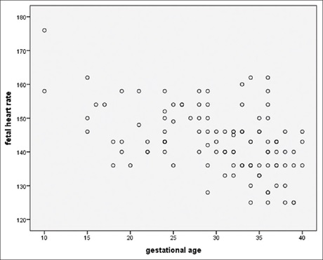
Results: The result obtained from the data was analyzed using Statistical Package for Social Sciences (SPSS) version 21. Mean, minimum, and maximum values were obtained. The relationship between the FHR, EFW, and GA was correlated. The minimum GA was n = 10 weeks while the maximum GA was n = 40 weeks with a mean value of 30. The minimum fetal weight was observed to be n = 35 g while the maximum fetal weight was n = 4402 g and the mean value was 1923.8 the minimum FHR obtained from this research was
n = 125 bpm and the maximum = 176 bpm while the mean was observed to be 143.4.
Conclusions: In summary, the FHR is affected by the EFW and the GA in such a manner that as the fetal weight and GA increase, the FHR decreases and vice versa. The FHR ranges from 125 bpm to 176 bpm in a healthy pregnant woman.
Downloads
Article Details
Section
This is an open access journal, and articles are distributed under the terms of the Creative Commons Attribution-NonCommercial-ShareAlike 4.0 License, which allows others to remix, tweak, and build upon the work non-commercially, as long as appropriate credit is given and the new creations are licensed under the identical terms.
How to Cite
References
1. Sontakke Y. Textbook of Human Embryology. London: CBS Publishers and Distributors; 2018. p. 180‑1.
2. Mohide P, Keirse MJ. Biophysical assessment of fetal wellbeing. In: Chalmers E, Enkin M, Kerise MJ, editors. Effective Care in Pregnancy and Child Birth. Oxford: Oxford University Press; 1991. p. 477‑92.
3. Hamelmann P, Vullings R, Kolen AF, Bergmans JW, van Laar JO, Tortoli P, et al. Doppler ultrasound technology for fetal heart rate monitoring: A review. IEEE Trans Ultrason Ferroelectr Freq Control 2020;67:226‑38.
4. Bhide A, Acharya G. Sex differences in fetal heart rate and variability assessed by antenatal computerized cardiotocography. Acta Obstet Gynecol Scand 2018;97:1486‑90.
5. Lim E, Miyamura J, Chen JJ. Racial/ethnic‑specific reference intervals for common laboratory tests: A comparison among Asians, blacks, hispanics, and white. Hawaii J Med Public Health 2015;74:302‑10.
6. Pildner von Steinburg S, Boulesteix AL, Lederer C, Grunow S, Schiermeier S, Hatzmann W, et al. What is the “normal” fetal heart
rate? PeerJ 2013;1:e82.
7. Avitan T, Sanders A, Brain U, Rurak D, Oberlander TF, Lim K. Variations from morning to afternoon of middle cerebral and umbilical artery blood flow, and fetal heart rate variability, and fetal characteristics in the normally developing fetus. J Clin Ultrasound 2018;46:235‑40.
8. Nigel B, Daniel P, Debbi S. Qualitative Methods for Health Research: A Practical Interactive Guide to Epidemiology and Statistics. Hoboken, New Jersey: John Wiley and Sons Limited; 2008. p. 145‑65.
9. Grivell RM, Alfirevic Z, Gyte GM, Devane D. Antenatal cardiotocography for fetal assessment. Cochrane Database Syst Rev 2015; 2015: CD007863.
10. Serra V, Bellver J, Moulden M, Redman CW. Computerized analysisof normal fetal heart rate pattern throughout gestation. Ultrasound Obstet Gynecol 2009;34:74‑9.
11. Sandman CA, Cordova CJ, Davis EP, Glynn LM, Buss C. Patterns of fetal heart rate response at ~30 weeks gestation predict size at birth.
J Dev Orig Health Dis 2011;2:212‑7.
12. Doubilet PM, Benson CB, Chow JS. Outcome of pregnancies with rapid embryonic heart rates in the early first trimester. AJR Am J
Roentgenol 2000;175:67‑9.
13. Nijhuis IJ, ten Hof J, Nijhuis JG, Mulder EJ, Narayan H, Taylor DJ, et al. Temporal organization of fetal behavior from 24‑weeks gestation onwards in normal and complicated pregnancies. Dev Psychobiol 1999;34:257‑68.
14. Afors K, Chandraharan E. Use of continuous electronic fetal monitoring in a preterm fetus: Clinical dilemmas and recommendations
for practice. J Pregnancy 2011;2011:848794.