

Pictorial Essay: A Retrospective Review of Male Breast Diseases in Maiduguri and Kano, Nigeria
Article Sidebar
Views | PDF/EPUB Downloads:
88
/ 10
/ 6
Main Article Content
Abstract
Introduction: Breast diseases in men are not as common as those in women and though male breast cancer is seen rarely, thus the
lack of screening guidelines worldwide, benign breast diseases such as gynecomastia present fairly commonly in both primary and
tertiary care setting. There is a paucity of information about the pattern, protocols, and imaging features of male breast diseases in
Nigeria.
Objective: To review the variety of presentations and radiological features of male breast diseases encountered in Aminu Kano
Teaching Hospital (AKTH) and University of Maiduguri Teaching Hospital (UMTH). We wish to discuss the departmental protocols and
highlight the role of mammography and sonomammography in the evaluation of male breast diseases.
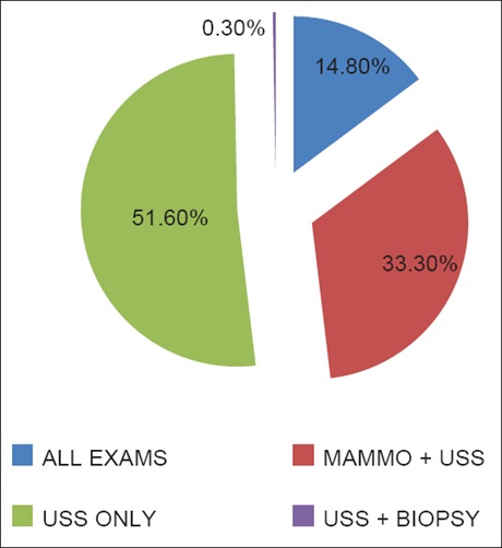
Materials and Methods: A 5‑year retrospective review was performed on the imaging findings of a total number of 27 male patients who presented with symptoms of breast disease to the radiology departments of AKTH (12) and UMTH (15) in Nigeria. All patients had mammography and sonomammography or sonomammography alone performed by a senior radiology resident and consultant radiologist. Selected cases had ultrasound guided biopsy and histology.
Results: Twenty‑seven male patients were reviewed from both centers with an age range of 0.06–69 years (mean of 33.11 ± 18.10 years). The majority of patients (88.9%) presented with breast enlargement only. Concerning laterality of disease, bilateral involvement was more common (59.3%). In unilateral disease, 33.3% of patients presented with left‑sided lesions while only 7.4% had right‑sided involvement. Gynecomastia was seen in twenty (20) patients and was the most common breast disease seen in male patients presenting for imaging in both centers. Breast abscesses were the second most common. We saw one case of bilateral male breast cancer. Overall, bilateral disease was far more common than unilateral.
Conclusion: Mammography is the most important first‑line imaging modality employed in the diagnosis of male breast diseases in our environment; sonomammography is an important and radiological modality of investigation used to differentiate gynecomastia from male breast cancer and breast abscess. Gynecomastia remains the most common occurring male breast disease in our study.
Downloads
Article Details
Section

This work is licensed under a Creative Commons Attribution-NonCommercial 4.0 International License.
This is an open access journal, and articles are distributed under the terms of the Creative Commons Attribution-NonCommercial-ShareAlike 4.0 License, which allows others to remix, tweak, and build upon the work non-commercially, as long as appropriate credit is given and the new creations are licensed under the identical terms.
How to Cite
References
1. Moore KL. Clinical Oriented Anatomy. 4th ed. Philadelphia, USA: Wolters Kluwer, Lippincott Williams and Wilkins; 1999.
2. Nguyen C, Kettler MD, Swirsky ME, Miller VI, Scott C, Krause R, et al. Male breast disease: Pictorial review with radiologic‑pathologic correlation. Radiographics 2013;33:763‑79.
3. Carrasco RM, Benito MA, Del Campo ER, Cordoba ES, Villejuif FR.Pictorial Review: Imaging Findings of Male Breast Lesions. Educational Exhibit. ECR; 2013.
4. Ryan S, McNicholas M, Eustace S. The breast. In: Anatomy for Diagnostic Imaging. 3rd ed. Philadelphia, USA: Saunders Elsevier; 2011. p. 313‑23.
5. Popli MB, Popli V, Bahl P, Solanki Y. Pictorial essay: Mammography of the male breast. Indian J Radiol Imaging 2009;19:278‑81.
6. Lattin GE Jr., Jesinger RA, Mattu R, Glassman LM. From the radiologic pathology archives: Diseases of the male breast: Radiologic‑pathologic correlation. Radiographics 2013;33:461‑89.
7. Appelbaum AH, Evans GF, Levy KR, Amirkhan RH, Schumpert TD. Mammographic appearances of male breast disease. Radiographics
1999;19:559‑68.
8. Dialani V, Baum J, Mehta TS. Sonographic features of gynecomastia. J Ultrasound Med 2010;29:539‑47.
9. Mustapha Z, Minoza K, Okedayo M, Ali AA, Nggada HA, Kyari M. An appraisal of male mammography in Maiduguri, North Eastern Nigeria. Borno Med J 2014;11:129‑33.
10. Berg WA, Yang WT. Special topics‑gynecomastia. In: Diagnostic Imaging: Breast. 2nd ed. Salt Lake City, UT 84106, Salt Lake City, Utah: Amirsys; 2014. p. 98‑103.
11. Stavros AT. Evaluation of the male breast. In: Breast Ultrasound. 1st ed. Philadelphia, USA: Lippincott Williams & Wilkins; 2004. p. 733.
12. Dickson G. Gynecomastia. Am Fam Physician 2012;85:716‑22. Available from: http://www.aafp.org/afp. [Last accessed on 2012 Apr 01].
13. McKiernan JF, Hull D. Breast development in the newborn. Arch Dis Child 1981;56:525‑9.
14. Charlot M, Béatrix O, Chateau F, Dubuisson J, Golfier F, Valette PJ, et al. Pathologies of the male breast. Diagn Interv Imaging 2013;94:26‑37.
15. Mahmood S, Sabih Z, Sabih D. Lymphoma presenting as gynaecomastia. Biomed Imaging Interv J 2011;7:e10.
16. Iuanow E, Kettler M, Slanetz PJ. Spectrum of disease in the male breast. AJR Am J Roentgenol 2011;196:W247‑59.
17. Johnson RE, Murad MH. Gynecomastia: Pathophysiology, evaluation, and management. Mayo Clin Proc 2009;84:1010‑5.
18. Fischer U, Baum F, Luftner‑Nagel S. Benign changes. In: Breast Imaging – Direct Diagnosis in Radiology. 1st ed. Stuttgart, Germany:
Thieme; 2007. p. 136‑8.
19. Chu KM, Chiu LF, Fung HS, Kwok KY, Wai AM, Siu JC, et al. An institutional audit and pictorial review of common male breast diseases. Hong Kong J Radiol 2011;14:15‑23.