

Candida Esophagitis: Feathery Appearance as a New Sign on Barium Esophagogram
Article Sidebar
Views | PDF/EPUB Downloads:
30
/ 5
/ 6
Main Article Content
Abstract
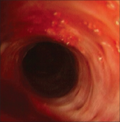
The characteristic appearance of Candida esophagitis on barium studies is that of diffuse discrete mucosal plaques, which may become confluent to form 'cobblestone or shaggy' esophagus. Many authors have also reported different radiographic findings such as a foamy appearance in florid esophageal candidiasis in immunocompromised patients. This report discusses a “feathery” appearance seen in barium esophagography of a 74‑year‑old woman who presented with dysphagia. The barium swallow showed fine out‑ pouching giving a “feathery” appearance, which is similar to what is described as pseudo‑diverticulosis in patients with esophagitis complicating gastro‑esophageal reflux disease. A diagnosis of esophagitis presumably due to candidiasis was made. This was confirmed by fungal studies on biopsy specimen following flexible esophagoscopy. Radiologists should be aware of this rare manifestation as a new sign of Candida esophagitis in order to avoid unnecessary delay in diagnosis and treatment.
Downloads
Article Details
Section

This work is licensed under a Creative Commons Attribution-NonCommercial 4.0 International License.
This is an open access journal, and articles are distributed under the terms of the Creative Commons Attribution-NonCommercial-ShareAlike 4.0 License, which allows others to remix, tweak, and build upon the work non-commercially, as long as appropriate credit is given and the new creations are licensed under the identical terms.
How to Cite
References
1. Levine MS, Rubesin SE. Diseases of the esophagus: Diagnosis with esophagography. Radiology 2005;237:414‑27.
2. Sam JW, Levine MS, Rubesin SE, Laufer I. The “foamy” esophagus: A radiographic sign of Candida esophagitis. AJR Am J Roentgenol 2000; 174:999‑1002.
3. Glick SN. Barium studies in patients with Candida esophagitis: Pseudoulcerations simulating viral esophagitis. AJR Am J Roentgenol 1994; 163:349‑52.
4. Gore RM, Levine MS. Textbook of Gastrointestinal Radiology. 2nd ed., Vol. 190. Philadelphia, PA: W.B. Sauders; 2000. p. 316‑509.
5. Al‑Shawwa B, D’Andrea L, Quintero D. Candida esophageal perforation and esophagopleural fistula: A case report. J Med Case Rep 2008; 2:209.
6. Levine MS, Macones AJ Jr, Laufer I. Candida esophagitis: Accuracy of radiographic diagnosis. Radiology 1985;154:581‑7.
7. Kumar P, Mohan S, Verma A, Baijal SS. Candida esophagitis in achalasia cardia: Case report and review of literature. Saudi J Gastroenterol 2007;13:88‑90.