

Department of Radiology, University of Ilorin Teaching Hospital, Ilorin, Nigeria
Article Sidebar
Views | PDF/EPUB Downloads:
74
/ 53
/ 19
Main Article Content
Abstract
The calcifying epithelial odontogenic cyst (CEOC) is a rare lesion of the jaws first described as a distinct entity by Gorlin et al., in 1962. [1] The condition is also referred as Gorlin’s cyst, keratinizing ameloblastoma or melanotic ameloblastic odontoma. [1] CEOC is often referred as an asymptomatic slow growing swelling of the jaws. It is a well circumscribed, solid or cystic lesion derived from odontogenic epithelium (OE) which develops from reduced enamel epithelium or remnants of OE in the follicle, gingival tissue or bone but contains “ghost cells” and spherical calcifications. It is considered a unique entity with both cystic and neoplastic behavior. [2] We report a case of CEOC which occurred in the maxillary sinus.
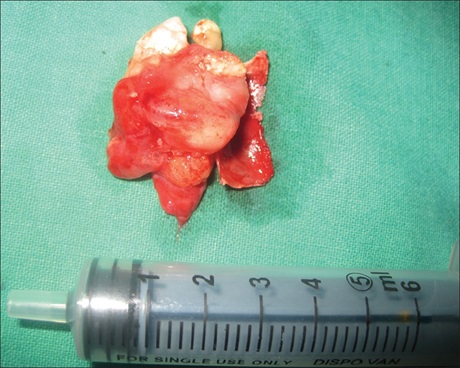
A 45-year-old female patient came with a chief complaint of asymptomatic swelling in the right upper jaw since 6 months. The lesion had been slowly increasing in size since it was first noticed. The lesion was extending from right lateral incisor to first molar on the same side intraorally and soft in consistency on palpation. A panoramic radiograph showed a well-circumscribed radiolucency in relation to upper right premolars with impacted canine and supernumerary tooth causing root resorption and displacing roots of premolars [Figure 1]. Computed tomography (CT) scan axial section revealed well-circumscribed radiolucency in the maxillary sinus thus obliterating the sinus [Figure 2], enucleation and aggressive curettage was done under local anesthesia using intraoral Caldwell-Luc approach [Figure 3]. Histopathology reviewed presence of cystic space lined by OE with ghost cells suggestive of CEOC [Figure 4]. Postoperative follow-up was done for 1 year and no recurrence was
observed.
Downloads
Article Details
Section

This work is licensed under a Creative Commons Attribution-NonCommercial 4.0 International License.
This is an open access journal, and articles are distributed under the terms of the Creative Commons Attribution-NonCommercial-ShareAlike 4.0 License, which allows others to remix, tweak, and build upon the work non-commercially, as long as appropriate credit is given and the new creations are licensed under the identical terms.
How to Cite
References
1. Gorlin RJ, Pindborg JJ, Odont, Clausen FP, Vickers RA. The calcifying odontogenic cyst‑a possible analogue of the cutaneous
calcifying epithelioma of Malherbe. An analysis of fifteen cases. Oral Surg Oral Med Oral Pathol 1962;15:1235‑43.
2. Freedman PD, Lumerrnan H, Gee JK. Calcifying odontogenic cyst. A review and analysis of seventy cases. Oral Surg Oral Med Oral
Pathol 1975;40:93‑106.
3. Praetorius FP. Calcifying odontogenic. Range, variation and neoplastic potential. Symposium on maxillofacial bone pathology.
Int J Oral Surg 1975;4:89.
4. Hirshberg A, Kaplan I, Buchner A. Calcifying odontogenic cyst associated with odontoma. A possible separate entity (odontocalcifying odontogenic cyst). J Oral Maxillofac Surg 1994;52:555‑8.
5. Balaji SM, Rooban T. Calcifying odontogenic cyst with atypical features. Ann Maxillofac Surg 2012;2:82‑5.
6. Hong SP, Ellis GL, Hartman KS. Calcifying odontogenic cyst. A review of ninety‑two cases with reevaluation of their nature as
cysts or neoplasms, the nature of ghost cells, and subclassification. Oral Surg Oral Med Oral Pathol 1991;72:56‑64.